. 2014 Jul;37(7):518-25.
doi: 10.14348/molcells.2014.0024. Epub 2014 Jun 30.

A gammaherpesvirus establishes persistent infection in neuroblastoma cells

Affiliations

A gammaherpesvirus establishes persistent infection in neuroblastoma cells

Hye-Jeong Cho et al. Mol Cells. 2014 Jul.

Abstract

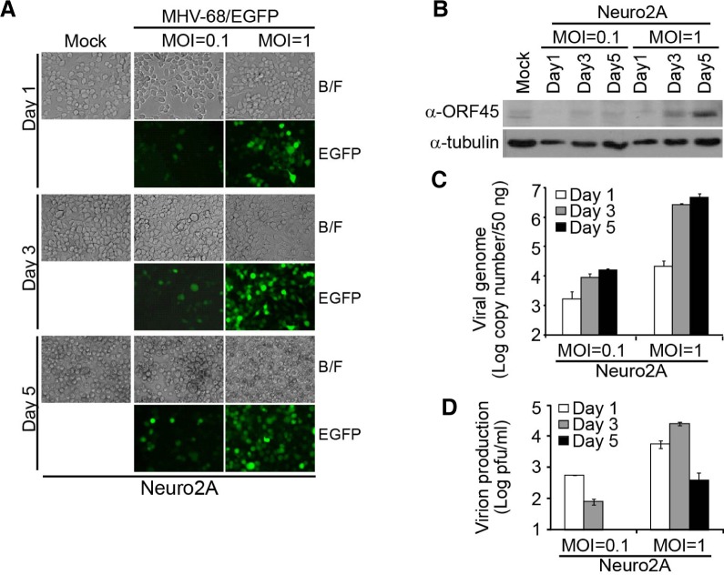
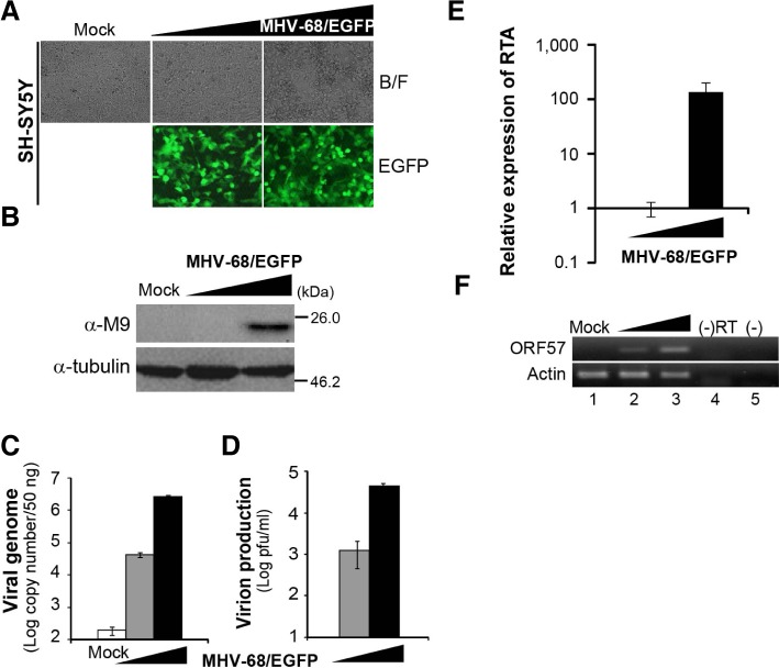
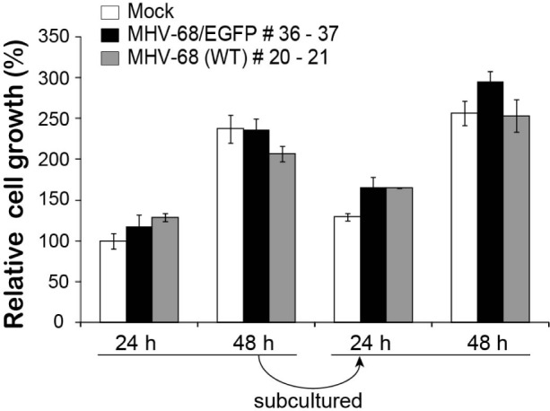
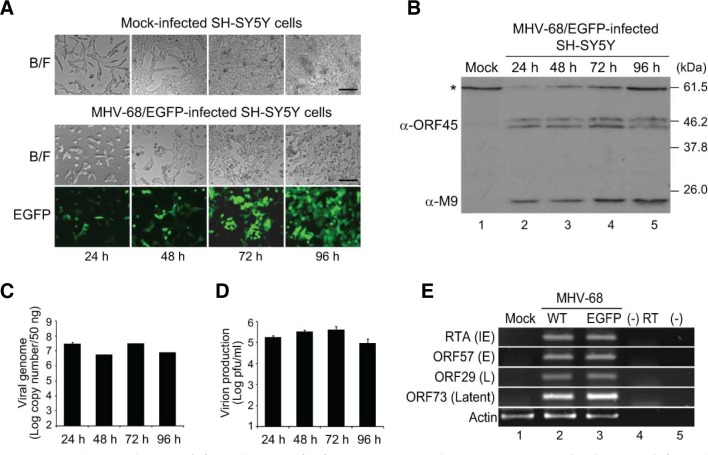
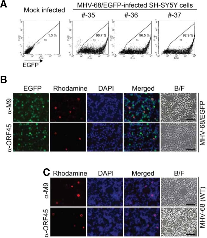
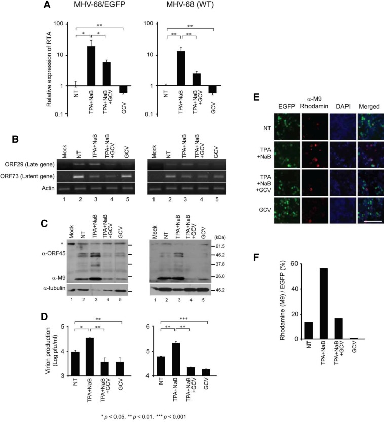
Gammaherpesvirus (γHV) infection of the central nervous system (CNS) has been implicated in diverse neurological diseases, and murine γHV-68 (MHV-68) is known to persist in the brain after cerebral infection. The underlying molecular mechanisms of persistency of virus in the brain are poorly understood. Here, we characterized a unique pattern of MHV-68 persistent infection in neuroblastoma cells. On infection with MHV-68, both murine and human neuroblastoma cells expressed viral lytic proteins and produced virions. However, the infected cells survived productive infection and could be cultured for multiple passages without affecting their cellular growth. Latent infection as well as productive replication was established in these prolonged cultures, and lytic replication was further increased by treatment with lytic inducers. Our results provide a novel system to study persistent infection of γHVs in vitro following de novo infection and suggest application of MHV-68 as a potential gene transfer vector to the brain.

Keywords: CNS; gammaherpesvirus; gene delivery; neuroblastoma; persistent infection.


Figures

Fig. 1.
Fig. 1.
Kinetic studies of MHV-68/EGFP infection in Neuro2A cells. Neuro2A, murine neuroblastoma cells, were infected with MHV-68/EGFP at an MOI of 0.1 or 1 and analyzed at 1, 3, and 5 days after infection for virus replication. (A) EGFP expression. The MHV-68/EGFP-infected and mock-infected Neuro2A cells were monitored for EGFP expression under a fluorescence microscope. The representative images are shown. B/F indicates the images from the bright field. (B) Viral lytic protein expression. The infected cell lysates were subjected to Western blotting with a polyclonal antibody against ORF45, a viral lytic protein. α-Tubulin was used as the loading control. (C) Replication of viral genomes. The copy numbers of viral genomic DNAs extracted from infected cells were measured by real-time PCR. Three independent assays were performed in duplicate, and the means ± standard deviation (SD) of the data are shown. (D) Production of infectious virions. The virus titers of the culture supernatants from the infected Neuro2A cells were measured by plaque assays. Three independent assays were performed, and the means ± SD of the data are shown. Mock-infected Neuro2A cells served as the negative control.
Fig. 2.
Fig. 2.
Non-cytolytic, productive infection of MHV-68 in SH-SY5Y cells. SH-SY5Y, human neuroblastoma cells, were infected with MHV-68/EGFP at an MOI of 0.1 or 1 for 72 h. Mock-infected SH-SY5Y cells were used as the negative control. EGFP expression (A), M9 viral lytic protein expression (B), viral genome replication (C), and virion production (D) from infected SH-SY5Y cells were analyzed as described in Fig. 1. Expression of an immediate early viral transcript, RTA, (E) and an early lytic transcript, ORF57, (F) were detected in SH-SY5Y cells by RT-qPCR and RT-PCR, respectively. Actin was used as an internal control for RT-qPCR and RT-PCR.
Fig. 3.
Fig. 3.
Effect of MHV-68 infection on the relative growth of SH-SY5Y cells. SH-SY5Y cells infected with MHV-68/EGFP or MHV-68 WT were subcultured for 36 passages or 20 passages, respectively. The infected cells were seeded in 96-well plates, cultured for 24 h and 48 h, and analyzed for cell viability by MTT assays. The cells were subcultured at 1:10 ratio and the cell viability was monitored for another 24 and 48 h. The relative cell growth was calculated based on the cell viability of mock-infected SH-SY5Y cells at 24 h after the first seeding. Three sets of samples were used in each experiment, and the experiments were independently performed twice. The means ± SD of the data are shown.
Fig. 4.
Fig. 4.
Persistent infection of MHV-68/EGFP infection in SH-SY5Y cells cultured for multiple passages. (A-D) MHV-68/EGFP-infected SH-SY5Y cells following 20 passages from the initial infection were monitored daily until 96 h after seeding. Expression of EGFP (A), viral lytic proteins, ORF45 and M9 (B), replication of viral genomic DNAs (C), and virion production (D) from prolonged infection of SH-SY5Y cells were analyzed as described in Fig. 1. The asterisk symbol indicates a non-specific band (B). Expression of viral lytic and latent transcripts (E). SH-SY5Y cells infected with MHV-68/EGFP or WT were cultured for 20 passages and harvested at 48 h post-seeding. Immediate-early (IE; RTA), early (E; ORF57), late (L; ORF29), and latent (ORF73) gene transcript were analyzed by RT-PCR. Actin was used as an internal control. Lanes 4 and 5 indicate negative controls without RT and template, respectively.
Fig. 5.
Fig. 5.
Both productive and latent replications of MHV-68/EGFP in prolonged culture of infected SH-SY5Y cells. (A) Persistent EGFP expression in the majority of MHV-68/EGFP-infected SH-SY5Y cells. MHV-68/EGFP-infected SH-SY5Y cells were grown for 35–37 passages from the initial infection and assayed by flow cytometry for EGFP expression. Mock-infected SH-SY5Y cells were used as the negative control. (B, C) Sporadic expression of lytic viral proteins in the infected SH-SY5Y cells. MHV-68/EGFP- or WT-infected SH-SY5Y cells were grown for 34 or 20 passages from the initial infection, respectively. Immunofluorescence assays were perfor-med using antibodies against ORF45 and M9 viral lytic proteins in the infected cells under a fluorescence microscope. The representative images are shown. B/F indicates the images from the bright field.
Fig. 6.
Fig. 6.
Induction of productive replication in prolonged culture of MHV-68/EGFP-infected SH-SY5Y cells. MHV-68/EGFP- and WT-infected SH-SY5Y cells were grown for 54 and 34 passages, respectively, and treated with TPA and NaB in the presence and absence of GCV for 36 h. (A) The relative expression of RTA transcript. The levels of RTA transcript measured by RT-qPCR. Actin was used as an internal control. The relative expression of the RTA transcript was calculated based on its level in the infected cells with no treatment (NT). (B) Expression of lytic and latent gene transcripts. The transcripts of a late gene (ORF29) and a latent gene (ORF73) were detected by RT-PCR. Actin was used as an internal control. (C) Expression of viral lytic proteins. The cell lysates were subjected to western blotting with antibodies against ORF45 and M9. Tubulin was used as the loading control; the asterisk symbol indicates the non-specific band. (D) Virion production. Virus titers were measured by plaque assays of the cultured supernatants harvested at 36 h after treatment. Statistical analysis was performed by Student’s t-test (A, D). (E–F) Expression of a lytic protein determined by immunofluorescence assay. The MHV-68/EGFP-infected cells were subjected to immunofluorescence assays with the antibody against M9, a small capsid protein. The representative images are shown. A minimum of 100 EGFP-positive cells were counted for each sample, and the percentage of M9-positive cells among the EGFP-positive cells was calculated and shown in a graphical form (F).

References

    1. Abrams Bae J, Lee D, Kim YK, Gil M, Lee JY, Lee KY. Berberine protects 6-hydroxydopamine-induced human dopaminergic neuronal cell death through the induction of heme oxygenase-1. Mol Cells. 2012;35:151–157. - PMC - PubMed
    1. Adler H, Messerle M, Koszinowski UH. Virus reconstituted from infectious bacterial artificial chromosome (BAC)-cloned murine gammaherpesvirus 68 acquires wild-type properties in vivo only after excision of BAC vector sequences. J Virol. 2001;75:5692–5696. - PMC - PubMed
    1. Bae J, Lee D, Kim YK, Gil M, Lee JY, Lee KY. Berberine protects 6-hydroxydopamine-induced human dopaminergic neuronal cell death through the induction of heme oxygenase-1. Mol Cells. 2012;35:151–157. - PMC - PubMed
    1. Bossolasco S, Falk KI, Ponzoni M, Ceserani N, Crippa F, Lazzarin A, Linde A, Cinque P. Ganciclovir is associated with low or undetectable Epstein-Barr virus DNA load in cerebrospinal fluid of patients with HIV-related primary central nervous system lymphoma. Clin Infect Dis. 2006;42:e21–25. - PubMed
    1. Cho HJ, Yu F, Sun R, Lee D, Song MJ. Lytic induction of Kaposi’s sarcoma-associated herpesvirus in primary effusion lymphoma cells with natural products identified by a cell-based fluorescence moderate-throughput screening. Arch Virol. 2008;153:1517–1525. - PubMed

Publication types

MeSH terms