Review
. 2014 Aug 4;206(3):335-45.
doi: 10.1083/jcb.201404154.

A unified cell biological perspective on axon-myelin injury

Affiliations
Review

A unified cell biological perspective on axon-myelin injury

Mikael Simons et al. J Cell Biol. .

Abstract

Demyelination and axon loss are pathological hallmarks of the neuroinflammatory disorder multiple sclerosis (MS). Although we have an increasingly detailed understanding of how immune cells can damage axons and myelin individually, we lack a unified view of how the axon-myelin unit as a whole is affected by immune-mediated attack. In this review, we propose that as a result of the tight cell biological interconnection of axons and myelin, damage to either can spread, which might convert a local inflammatory disease process early in MS into the global progressive disorder seen during later stages. This mode of spreading could also apply to other neurological disorders.


Figures

Figure 1.
Figure 1.
Molecular composition of the axon–myelin unit. Schematic diagram and electron microscopy images showing the different domains of a myelinated axon. Each of these domains is characterized by the expression of specific proteins: the node of Ranvier contains voltage-gated Na+ channels (NaCh), the paranode is the region where myelin is tightly attached to the axon by a complex formed by neurofascin-155 (NF155), contactin-1 (Cntn1), and contactin-associated protein (Caspr1), and the juxtaparanode where most voltage-gated K+ channels (KCh) are found is separated from the paranodes by a complex formed by contactin-associated protein 2 (Caspr2) and contactin-2 (Cntn2). The internodal region contains several cell adhesion molecules (Nectin-like molecule 1 [Necl1], Nectin-like molecule 4 [Necl4], and myelin-associated glycoprotein [Mag]). Bars, 80 nm.
Figure 2.
Figure 2.
Patterns of axon and myelin injury. (A, left) Axonal damage can either follow a classical centrifugal Wallerian pattern emanating from the cell body or an axonal transection. (middle) In contrast, an axonal die-back pattern originates at or near the synaptic compartment and spreads centripetally. (right) Finally, focal forms of axon injury have been described, e.g., during neuroinflammatory attack. (B) Similarly, in oligodendrocytes, injury can start at the soma (left), at the myelin sheaths (oligodendrocytic die back; middle), or focally at a single myelin-bearing process (right).
Figure 3.
Figure 3.
Mechanisms of coupling injury within the axon–myelin unit. (A–C) Proposed mechanisms of injury that affect the axon–myelin unit involve several nonexclusive scenarios, including loss of trophic or metabolic support (A), increased energy demand as a result of loss of myelin or compromised membranes (B), and gain of toxic functions, either within the axon or emanating from the myelin (C). P, phosphorylation.
Figure 4.
Figure 4.
Topologic similarities between neurons and oligodendrocytes. (A and B) As extended, process-bearing cells, neurons (A) and oligodendrocytes (B) show similarities in their topology, including a trophic center, the soma, which is connected with a metabolically highly active periphery (dark red; synapses for neurons and the inner myelin tongue for oligodendrocytes) through a rather thin and vulnerable conduit (a neuron’s axon and oligodendrocytic processes, respectively). Oligodendrocytes carry a large additional membrane compartment, compacted myelin (teal)—which in the main drawing is shown in a hypothetical “unrolled” state and also schematized in the cross section. Arrowheads point to the axon (A) and an oligodendrocyte process (B).
Figure 5.
Figure 5.
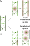
Mechanisms of spread of axon–myelin injury. (A and B) The axon–myelin unit imposes a tight coupling of neuronal and oligodendrocytic health. This could result in different patterns of spreading pathology—e.g., a transversal pattern (A), in which primary loss of an oligodendrocyte injures all of the axons it subserves, versus a longitudinal pattern (B), in which Wallerian degeneration affects all oligodendrocytes distal from a primary axonal lesion. Gray, axons; green, oligodendrocytes; red haze, site of injury.

References

    1. Aboul-Enein, F., Rauschka H., Kornek B., Stadelmann C., Stefferl A., Brück W., Lucchinetti C., Schmidbauer M., Jellinger K., and Lassmann H.. 2003. Preferential loss of myelin-associated glycoprotein reflects hypoxia-like white matter damage in stroke and inflammatory brain diseases. J. Neuropathol. Exp. Neurol. 62:25–33 - PubMed
    1. Aggarwal, S., Yurlova L., and Simons M.. 2011. Central nervous system myelin: structure, synthesis and assembly. Trends Cell Biol. 21:585–593 10.1016/j.tcb.2011.06.004 - DOI - PubMed
    1. Aggarwal, S., Snaidero N., Pähler G., Frey S., Sánchez P., Zweckstetter M., Janshoff A., Schneider A., Weil M.T., Schaap I.A., et al. . 2013. Myelin membrane assembly is driven by a phase transition of myelin basic proteins into a cohesive protein meshwork. PLoS Biol. 11:e1001577 10.1371/journal.pbio.1001577 - DOI - PMC - PubMed
    1. Arancibia-Carcamo, I.L., and Attwell D.. 2014. The node of Ranvier in CNS pathology. Acta Neuropathol. 10.1007/s00401-014-1305-z - DOI - PMC - PubMed
    1. Arthur-Farraj, P., Mirsky R., Parkinson D.B., and Jessen K.R.. 2006. A double point mutation in the DNA-binding region of Egr2 switches its function from inhibition to induction of proliferation: A potential contribution to the development of congenital hypomyelinating neuropathy. Neurobiol. Dis. 24:159–169 10.1016/j.nbd.2006.06.006 - DOI - PubMed

Publication types