. 2014 Aug 5;9(8):e104330.
doi: 10.1371/journal.pone.0104330. eCollection 2014.

Kisspeptins modulate the biology of multiple populations of gonadotropin-releasing hormone neurons during embryogenesis and adulthood in zebrafish (Danio rerio)

Affiliations

Kisspeptins modulate the biology of multiple populations of gonadotropin-releasing hormone neurons during embryogenesis and adulthood in zebrafish (Danio rerio)

Yali Zhao et al. PLoS One. .

Abstract

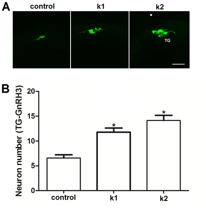
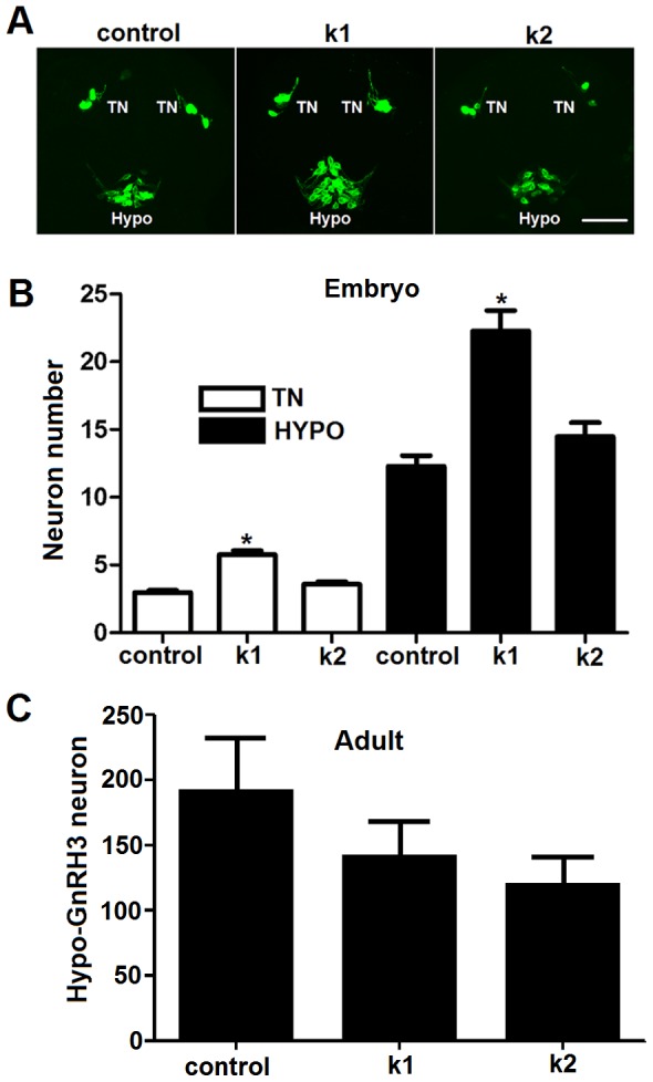
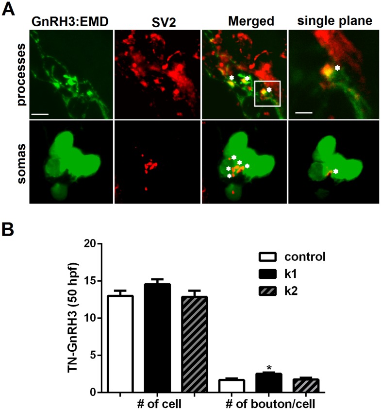
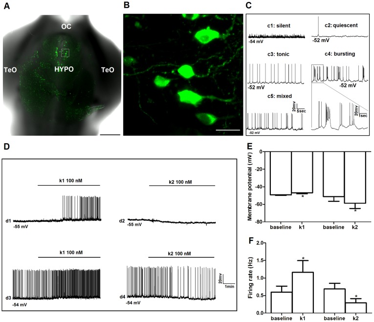
Kisspeptin1 (product of the Kiss1 gene) is the key neuropeptide that gates puberty and maintains fertility by regulating the gonadotropin-releasing hormone (GnRH) neuronal system in mammals. Inactivating mutations in Kiss1 and the kisspeptin receptor (GPR54/Kiss1r) are associated with pubertal failure and infertility. Kiss2, a paralogous gene for kiss1, has been recently identified in several vertebrates including zebrafish. Using our transgenic zebrafish model system in which the GnRH3 promoter drives expression of emerald green fluorescent protein, we investigated the effects of kisspeptins on development of the GnRH neuronal system during embryogenesis and on electrical activity during adulthood. Quantitative PCR showed detectable levels of kiss1 and kiss2 mRNA by 1 day post fertilization, increasing throughout embryonic and larval development. Early treatment with Kiss1 or Kiss2 showed that both kisspeptins stimulated proliferation of trigeminal GnRH3 neurons located in the peripheral nervous system. However, only Kiss1, but not Kiss2, stimulated proliferation of terminal nerve and hypothalamic populations of GnRH3 neurons in the central nervous system. Immunohistochemical analysis of synaptic vesicle protein 2 suggested that Kiss1, but not Kiss2, increased synaptic contacts on the cell body and along the terminal nerve-GnRH3 neuronal processes during embryogenesis. In intact brain of adult zebrafish, whole-cell patch clamp recordings of GnRH3 neurons from the preoptic area and hypothalamus revealed opposite effects of Kiss1 and Kiss2 on spontaneous action potential firing frequency and membrane potential. Kiss1 increased spike frequency and depolarized membrane potential, whereas Kiss2 suppressed spike frequency and hyperpolarized membrane potential. We conclude that in zebrafish, Kiss1 is the primary stimulator of GnRH3 neuronal development in the embryo and an activator of stimulating hypophysiotropic neuron activities in the adult, while Kiss2 plays an additional role in stimulating embryonic development of the trigeminal neuronal population, but is an RFamide that inhibits electrical activity of hypophysiotropic GnRH3 neurons in the adult.


Conflict of interest statement

Competing Interests: The authors have declared that no competing interests exist.

Figures

Figure 1
Figure 1. Levels of kiss1and kiss2 mRNAs in embryonic and larval zebrafish (1–7 days post-fertilization (dpf).
Quantitative RT-PCR values were normalized against elongation factor 1a and represent fold changes of kiss1 mRNA (top panel) and kiss2 mRNA (bottom panel) relative to that of 1 dpf embryos (n = 4 replicate experiments). Different letters denote significant differences between groups (at least P<0.05).
Figure 2
Figure 2. Kiss1 and Kiss2 stimulate proliferation of trigeminal GnRH3 neurons.
(A) Confocal images of lateral view of trigeminal (TG) GnRH3:EMD neurons at 18 h post fertilization (hpf). Scale bar, 50 µm. (B) Both Kiss1 (k1) and Kiss2 (k2) increased the number of TG-GnRH3:EMD neurons compared to controls at 18 hpf (at least n = 7 embryos per group). *: p<0.05 compared to controls.
Figure 3
Figure 3. Kiss1 but not Kiss2 stimulates proliferation of terminal nerve and hypothalamic GnRH3 neurons in embryos.
(A) Confocal images of ventral view showing the expression of TN-GnRH3:EMD neurons (TN) and hypothalamic GnRH3:EMD neurons (HYPO) at 25 hpf of a representative control embryo, and embryos treated with Kiss1 (k1) and Kiss2 (k2). Scale bar, A: 50 µm. (B) Summary data of the number of neurons expressed at 25 hpf in TN and HYPO (at least n = 14 embryos per group). Kiss1, but not Kiss2, significantly increased GnRH3 neuron numbers in both terminal nerve and hypothalamus. *: p<0.05 compared to controls. (C) Summary data of the number of hypothalamic GnRH3:EMD neurons in adult brains of zebrafish treated as embryos with 100 µM Kiss1 (n = 5), 100 µM Kiss2 (n = 4) or control solution (n = 5) (treatment duration: 5 hpf - 3 dpf).
Figure 4
Figure 4. Effects of Kiss1 and Kiss2 treatments on bouton formation on terminal nerve (TN)-GnRH3 neurons.
(A) Representative images of a TN-GnRH3:EMD neuron cluster in a 50 hpf embryo from the Control group, showing synaptic boutons in the neuronal processes (upper panels) and somas (lower panels) labeled with antibody to the presynaptic marker SV2. Far left panels: EMD expression in GnRH3 neurons (green); Middle left panels: Immunoreactive SV2 (red); Middle right panels: merged images of EMD and SV2 showing synaptic boutons in yellow; Far right panels: upper image: single plane (1 µm optical slice) with higher magnification of the boxed area in the upper middle right panel showing the synaptic contacts in the neuronal processes; lower image: single plane (1 µm optical slice) showing a synaptic bouton on the soma. Each asterisk labels a nearby bouton. (B) Total number of TN-GnRH3:EMD neurons in the bilateral clusters (left), and number of boutons normalized to the total number of neurons in the clusters (soma and processes; right). Kiss1 and Kiss2 had no effect on the number of TN-GnRH3 neurons at 50 hpf. However, Kiss1, but not Kiss2, significantly increased the number of boutons per TN-GnRH3 neuron (n = 7 embryos per group). This increase was seen in both cell bodies and neural projections. *: p<0.05 compared to controls. Scale bars: 5 µm for all images, except the one in the upper far right panel where it is 2 µm.
Figure 5
Figure 5. Effects of Kiss1 and Kiss2 on electrical activity of hypophysiotropic GnRH3:EMD neurons in adult zebrafish brain.
(A) Low power confocal image of ventral view of excised, intact brain showing GnRH3:EMD neurons expressed in hypothalamus. OC: optic chiasm; HYPO: hypothalamus; TeO: optic tectum. Scale bar, 200 µm. (B) High magnification confocal image of the white boxed region in (A). Scale bar, 10 µm. (C) Sample electrophysiology traces of different patterns of spontaneous action potential firing from GnRH3:EMD neurons located in the preoptic area and hypothalamus of the intact adult brain: silent (c1); quiescent (c2); tonic (c3); bursting (c4); mixed tonic and bursting (c5). The boxed area in c4 is expanded to show details of the firing pattern. (D) Sample traces from four neurons showing the responses to Kiss1 (k1: d1 and d3) and Kiss2 (k2: d2 and d4), with 2 min baseline and 5 min treatment period. d1 and d2: silent neurons; d3 and d4: active neurons. Summary of the effects of k1 and k2 treatments on membrane potential (E) and spike frequency (F). *: p<0.05 compared to baseline values. At least n = 6 neurons for each treatment group.

References

    1. Funes S, Hedrick JA, Vassileva G, Markowitz L, Abbondanzo S, et al. (2003) The KiSS-1 receptor GPR54 is essential for the development of the murine reproductive system. Biochem Biophys Res Commun 312: 1357–1363. - PubMed
    1. Clarkson J, d'Anglemont de Tassigny X, Moreno AS, Colledge WH, Herbison AE (2008) Kisspeptin-GPR54 signaling is essential for preovulatory gonadotropin-releasing hormone neuron activation and the luteinizing hormone surge. J Neurosci 28: 8691–8697. - PMC - PubMed
    1. Oakley AE, Clifton DK, Steiner RA (2009) Kisspeptin signaling in the brain. Endocr Rev 30: 713–743. - PMC - PubMed
    1. Seminara SB, Messager S, Chatzidaki EE, Thresher RR, Acierno JS Jr, et al. (2003) The GPR54 gene as a regulator of puberty. N Engl J Med 349: 1614–1627. - PubMed
    1. Oka Y (2009) Three types of gonadotrophin-releasing hormone neurones and steroid-sensitive sexually dimorphic kisspeptin neurones in teleosts. J Neuroendocrinol 21: 334–338. - PubMed

Publication types

MeSH terms