

Axon diameter and intra-axonal volume fraction of the corticospinal tract in idiopathic normal pressure hydrocephalus measured by q-space imaging
- PMID: 25093733
- PMCID: PMC4122461
- DOI: 10.1371/journal.pone.0103842
Axon diameter and intra-axonal volume fraction of the corticospinal tract in idiopathic normal pressure hydrocephalus measured by q-space imaging
Abstract
Purpose: Previous studies suggest that compression and stretching of the corticospinal tract (CST) potentially cause treatable gait disturbance in patients with idiopathic normal pressure hydrocephalus (iNPH). Measurement of axon diameter with diffusion MRI has recently been used to investigate microstructural alterations in neurological diseases. In this study, we investigated alterations in the axon diameter and intra-axonal fraction of the CST in iNPH by q-space imaging (QSI) analysis.
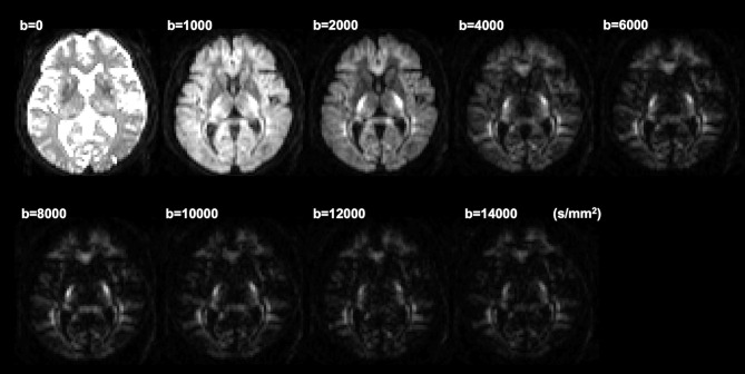
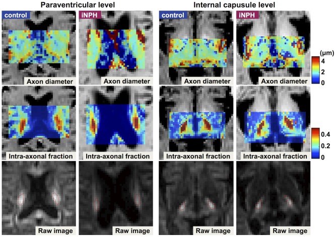
Methods: Nineteen patients with iNPH and 10 age-matched controls were recruited. QSI data were obtained with a 3-T system by using a single-shot echo planar imaging sequence with the diffusion gradient applied parallel to the antero-posterior axis. By using a two-component low-q fit model, the root mean square displacements of intra-axonal space ( = axon diameter) and intra-axonal volume fraction of the CST were calculated at the levels of the internal capsule and body of the lateral ventricle, respectively.
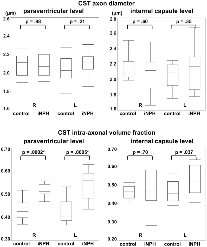
Results: Wilcoxon's rank-sum test revealed a significant increase in CST intra-axonal volume fraction at the paraventricular level in patients (p<0.001), whereas no significant difference was observed in the axon diameter. At the level of the internal capsule, neither axon diameter nor intra-axonal volume fraction differed significantly between the two groups.
Conclusion: Our results suggest that in patients with iNPH, the CST does not undergo irreversible axonal damage but is rather compressed and/or stretched owing to pressure from the enlarged ventricle. These analyses of axon diameter and intra-axonal fraction yield insights into microstructural alterations of the CST in iNPH.
Conflict of interest statement
Figures
Similar articles
-
Prospective estimation of mean axon diameter and extra-axonal space of the posterior limb of the internal capsule in patients with idiopathic normal pressure hydrocephalus before and after a lumboperitoneal shunt by using q-space diffusion MRI.Eur Radiol. 2016 Sep;26(9):2992-8. doi: 10.1007/s00330-015-4162-9. Epub 2015 Dec 22. Eur Radiol. 2016. PMID: 26694062 Free PMC article.
-
Diffusion imaging of reversible and irreversible microstructural changes within the corticospinal tract in idiopathic normal pressure hydrocephalus.Neuroimage Clin. 2017 Mar 11;14:663-671. doi: 10.1016/j.nicl.2017.03.003. eCollection 2017. Neuroimage Clin. 2017. PMID: 28348958 Free PMC article.
-
Microstructural changes of the corticospinal tract in idiopathic normal pressure hydrocephalus: a comparison of diffusion tensor and diffusional kurtosis imaging.Neuroradiology. 2013 Aug;55(8):971-976. doi: 10.1007/s00234-013-1201-6. Epub 2013 Jun 2. Neuroradiology. 2013. PMID: 23728069
-
The role of diffusion tensor imaging and fractional anisotropy in the evaluation of patients with idiopathic normal pressure hydrocephalus: a literature review.Neurosurg Focus. 2016 Sep;41(3):E12. doi: 10.3171/2016.6.FOCUS16192. Neurosurg Focus. 2016. PMID: 27581308 Review.
-
[Current State of Diagnosis and Treatment of Idiopathic Normal Pressure Hydrocephalus].Brain Nerve. 2016 Apr;68(4):429-40. doi: 10.11477/mf.1416200416. Brain Nerve. 2016. PMID: 27056861 Review. Japanese.
Cited by
-
Advanced diffusion-weighted magnetic resonance imaging in the evaluation of white matter axons in patients with idiopathic normal pressure hydrocephalus.Neural Regen Res. 2017 Dec;12(12):1974-1975. doi: 10.4103/1673-5374.221149. Neural Regen Res. 2017. PMID: 29323031 Free PMC article. No abstract available.
-
Model-Based Comparison of Deep Brain Stimulation Array Functionality with Varying Number of Radial Electrodes and Machine Learning Feature Sets.Front Comput Neurosci. 2016 Jun 10;10:58. doi: 10.3389/fncom.2016.00058. eCollection 2016. Front Comput Neurosci. 2016. PMID: 27375470 Free PMC article.
-
Extra-axonal contribution to double diffusion encoding-based pore size estimates in the corticospinal tract.MAGMA. 2023 Aug;36(4):589-612. doi: 10.1007/s10334-022-01058-8. Epub 2023 Feb 6. MAGMA. 2023. PMID: 36745290 Free PMC article.
-
Prospective estimation of mean axon diameter and extra-axonal space of the posterior limb of the internal capsule in patients with idiopathic normal pressure hydrocephalus before and after a lumboperitoneal shunt by using q-space diffusion MRI.Eur Radiol. 2016 Sep;26(9):2992-8. doi: 10.1007/s00330-015-4162-9. Epub 2015 Dec 22. Eur Radiol. 2016. PMID: 26694062 Free PMC article.
-
MR Elastography Demonstrates Increased Brain Stiffness in Normal Pressure Hydrocephalus.AJNR Am J Neuroradiol. 2016 Mar;37(3):462-7. doi: 10.3174/ajnr.A4560. Epub 2015 Nov 5. AJNR Am J Neuroradiol. 2016. PMID: 26542235 Free PMC article.
References
-
- Relkin N, Marmarou A, Klinge P, Bergsneider M, Black PM (2005) Diagnosing idiopathic normal-pressure hydrocephalus. Neurosurgery 57: : S4–16; discussion ii–v. - PubMed
-
- Sasaki M, Honda S, Yuasa T, Iwamura A, Shibata E, et al. (2008) Narrow CSF space at high convexity and high midline areas in idiopathic normal pressure hydrocephalus detected by axial and coronal MRI. Neuroradiology 50: 117–122. - PubMed
-
- Marmarou A, Young HF, Aygok GA, Sawauchi S, Tsuji O, et al. (2005) Diagnosis and management of idiopathic normal-pressure hydrocephalus: a prospective study in 151 patients. Journal of Neurosurgery 102: 987–997. - PubMed
-
- Meier U, Lemcke J (2006) Clinical outcome of patients with idiopathic normal pressure hydrocephalus three years after shunt implantation. Acta Neurochir Suppl 96: 377–380. - PubMed
-
- Hakim S, Venegas JG, Burton JD (1976) The physics of the cranial cavity, hydrocephalus and normal pressure hydrocephalus: mechanical interpretation and mathematical model. Surgical Neurology 5: 187–210. - PubMed
Publication types
MeSH terms
LinkOut - more resources
Full Text Sources
Other Literature Sources
