Review
. 2014 Aug;32(8):804-18.
doi: 10.1038/nbt.2993.

Clinical imaging in regenerative medicine

Affiliations
Review

Clinical imaging in regenerative medicine

Anna V Naumova et al. Nat Biotechnol. 2014 Aug.

Abstract

In regenerative medicine, clinical imaging is indispensable for characterizing damaged tissue and for measuring the safety and efficacy of therapy. However, the ability to track the fate and function of transplanted cells with current technologies is limited. Exogenous contrast labels such as nanoparticles give a strong signal in the short term but are unreliable long term. Genetically encoded labels are good both short- and long-term in animals, but in the human setting they raise regulatory issues related to the safety of genomic integration and potential immunogenicity of reporter proteins. Imaging studies in brain, heart and islets share a common set of challenges, including developing novel labeling approaches to improve detection thresholds and early delineation of toxicity and function. Key areas for future research include addressing safety concerns associated with genetic labels and developing methods to follow cell survival, differentiation and integration with host tissue. Imaging may bridge the gap between cell therapies and health outcomes by elucidating mechanisms of action through longitudinal monitoring.


Conflict of interest statement

COMPETING FINANCIAL INTERESTS

The authors declare competing financial interests: details are available in the online version of the paper.

Figures

Figure 1
Figure 1
The human body contains ~3.7 × 10¹³ cells. The cell number and weights of individual organs, provide a baseline for understanding the numbers of cells that may be needed for replacement therapies and the associated challenges for imaging.
Figure 2
Figure 2
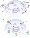
Tracking cell fate by noninvasive imaging requires either direct or indirect labeling. (a) Direct labeling. Exogenous labeling with either MRI-based contrast agents or radioprobes for PET or SPECT imaging. Cells take up SPIO or 19F nanoparticles (Fnp) primarily through endocytosis, whereas 99mTcHMPAO or 111Indium oxine are lipophilic and pass through cell membranes by passive diffusion. FDG is a glucose analog and is taken up though glucose transporter channels on cells. Small molecules can attach to cell surface markers or enter cells through channels. (b) Indirect labeling. Reporter genes introduced into the genome express surface proteins, channels, storage proteins or enzymes that are detectable or that bind detectable probes. HSV1-tk, herpes simplex virus thymidine kinase; NIS, sodium iodide symporter; LRP, lysine-rich protein; SPIO, superparamagnetic iron oxide nanoparticles.
Figure 3
Figure 3
Examples of clinical imaging used to identify and track labeled cells in the body. (a) Coronal SPECT images of chest and upper abdomen obtained 18 h after intracoronary (IC) versus transendocardial (TE) delivery of about 108 99mTcHMPAO CD34+ hematopoietic stem cells in patients with nonischemic dilated cardiomyopathy. IC route of administration clearly shows less retention of cells in the myocardium compared to TE route with both delivery techniques showing the distribution of CD34+ cells in liver and spleen (IC > TE). This comparison demonstrates the value of short-term labeling of a cell product and the value in assessing the cell delivery route. Figure from ref. reprinted with permission. (b) PET/CT fused axial multimodal imaging of pig chest performed on clinical scanner. MSC transduced by adenovirus containing cytomegalovirus promoter driving HSV1-tk reporter gene implanted with matrigel into porcine left ventricle (LV) myocardium after thoracotomy (long arrows). T is the chest tube inserted during surgery. 108 human MSCs injected into the myocardium were visualized (short arrows) 4 h after intravenous infusion of 9-(4-18F-3-[hydroxymethyl]butyl)-guanine, a thymidine analog that is phosphorylated by HSV1-tk reporter following uptake by cells. % ID/g is percentage uptake per gram of tissue. Figure from ref. reprinted with permission. (c) Coronal MRI performed at 3 T of liver from a diabetic patient baseline, day 1 and day 7 following portal vein infusion of 4.81 × 10⁵SPIO-labeled islet equivalents demonstrating hypointense voxels throughout the liver (white arrows) that cleared rapidly over 24 weeks of imaging follow-up. The number of hypointense areas in liver decreased by about 50–60% in the first week after infusion, representing rapid clearance of the transplanted islets despite immunosuppression of the patient. Figure adapted from ref. with permission. (d) Coronal and axial, T2-weighted, 1.0 T MRI following implantation of fetal neurons in a patient with Parkinson’s disease. Coronal MRI displays the cell transplantation needle track (arrowheads) following injection of ~3.2 × 10⁶ cells. 18F fluorodopa PET parametric map fused with MRI performed before (PRE) and 3 years post implantation (PI) of fetal cell neurons that matured with time that resulted in increased uptake of the dopamine analog by the innervated cells in the putamen. Figure adapted from ref. with permission. (e) T2-weighted, axial, 3 T MRI before (PRE) and day 1 after implantation in the left temporal lobe of SPIO-labeled autologous neural stem cells in patient with traumatic brain injury (* is site of injury). Magnified serial T2*-weighted, axial MRI performed on days (D) 1,7,14,21 post implantation (PI) of labeled cells are shown. Four hypointense areas (black arrows) were observed on PI days 1,7,14, 21 around lesion site (*) followed by migration of cells (white arrowhead and arrows) along the border of the damaged area. Hypointense areas where labeled cells were injected cleared over time (D14, D21), and by week 7 PI, areas were no longer visible on MRI. Figure adapted from ref. with permission.

Comment in

  • Cardiac regeneration validated.
    Malliaras K, Marbán E. Malliaras K, et al. Nat Biotechnol. 2015 Jun;33(6):587. doi: 10.1038/nbt.3254. Nat Biotechnol. 2015. PMID: 26057969 No abstract available.
  • Response to cardiac regeneration validated.
    Naumova AV, Modo M, Moore A, Frank JA, Murry CE. Naumova AV, et al. Nat Biotechnol. 2015 Jun;33(6):587. doi: 10.1038/nbt.3257. Nat Biotechnol. 2015. PMID: 26057970 No abstract available.

Similar articles

Cited by

References

    1. Daar AS, Greenwood HL. A proposed definition of regenerative medicine. J Tissue Eng Regen Med. 2007;1:179–184. - PubMed
    1. Robey TE, Saiget MK, Reinecke H, Murry CE. Systems approaches to preventing transplanted cell death in cardiac repair. J Mol Cell Cardiol. 2008;45:567–581. - PMC - PubMed
    1. de Almeida PE, van Rappard JR, Wu JC. In vivo bioluminescence for tracking cell fate and function. Am J Physiol Heart Circ Physiol. 2011;301:H663–H671. - PMC - PubMed
    1. Nguyen PK, Riegler J, Wu JC. Stem cell imaging: from bench to bedside. Cell Stem Cell. 2014;14:431–444. - PMC - PubMed
    1. Roura S, Galvez-Monton C, Bayes-Genis A. Bioluminescence imaging: a shining future for cardiac regeneration. J Cell Mol Med. 2013;17:693–703. - PMC - PubMed