. 2015 Jan;34(1):61-71.
doi: 10.1109/TMI.2014.2343916. Epub 2014 Jul 30.

PET image reconstruction using kernel method

PET image reconstruction using kernel method

Guobao Wang et al. IEEE Trans Med Imaging. 2015 Jan.

Abstract

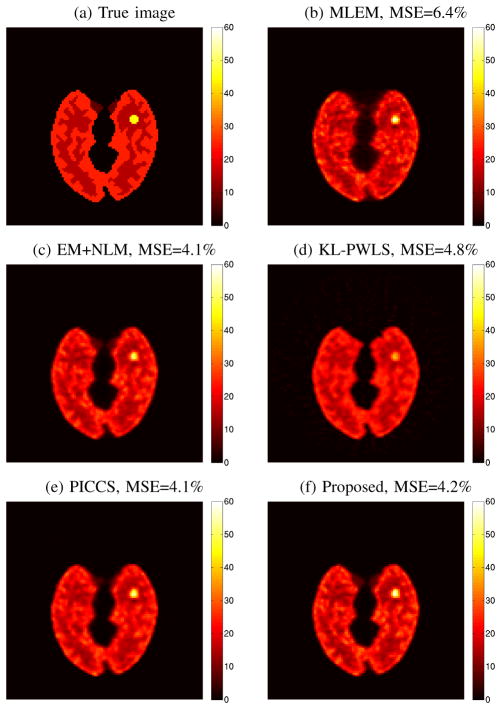
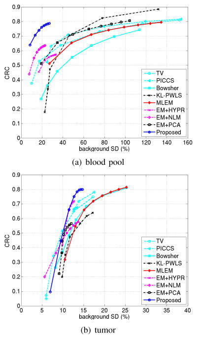
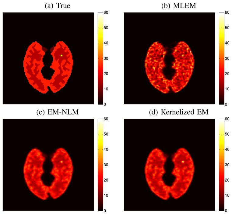
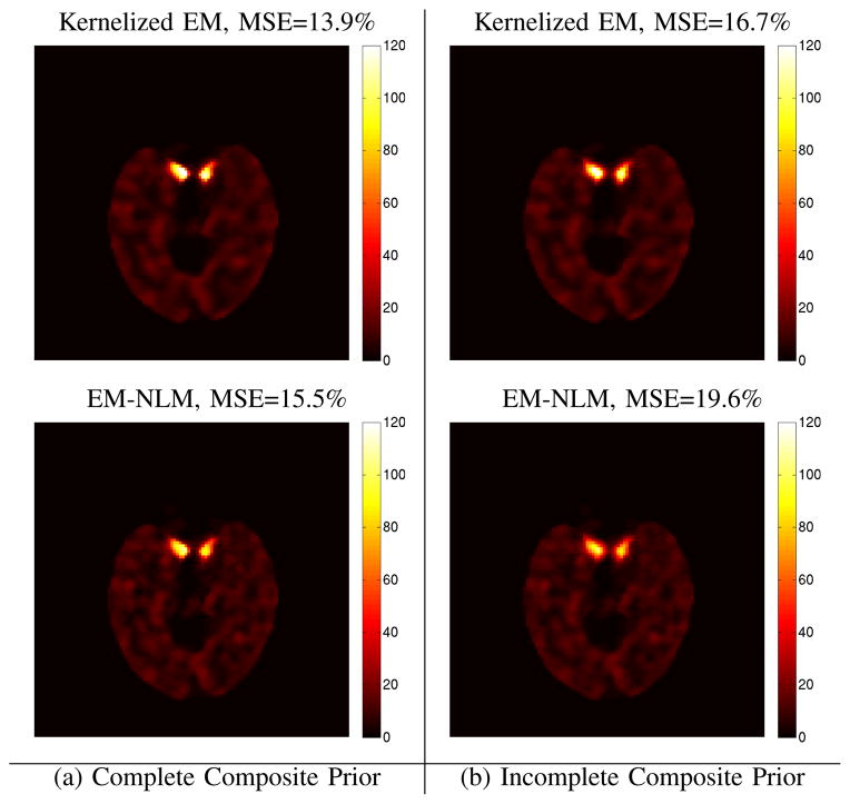
Image reconstruction from low-count positron emission tomography (PET) projection data is challenging because the inverse problem is ill-posed. Prior information can be used to improve image quality. Inspired by the kernel methods in machine learning, this paper proposes a kernel based method that models PET image intensity in each pixel as a function of a set of features obtained from prior information. The kernel-based image model is incorporated into the forward model of PET projection data and the coefficients can be readily estimated by the maximum likelihood (ML) or penalized likelihood image reconstruction. A kernelized expectation-maximization algorithm is presented to obtain the ML estimate. Computer simulations show that the proposed approach can achieve better bias versus variance trade-off and higher contrast recovery for dynamic PET image reconstruction than the conventional maximum likelihood method with and without post-reconstruction denoising. Compared with other regularization-based methods, the kernel method is easier to implement and provides better image quality for low-count data. Application of the proposed kernel method to a 4-D dynamic PET patient dataset showed promising results.


Figures

Fig. 1
Fig. 1
The digital phantom and time activity curves used in the simulation studies. (a) The Zubal brain phantom composed of gray matter, white matter and a tumor (15mm in diameter); (b) the regional time activity curves.
Fig. 2
Fig. 2
Composite images reconstructed from the rebinned dynamic sinograms: (a) the first 20 minutes, (b) the middle 20 minutes, and (c) the last 20 minutes.
Fig. 3
Fig. 3
Basis images reshaped from four different columns of the kernel matrix derived from the composite images shown in Fig. 2. The four chosen pixels are located in the blood region, at the gray matter edge, in the white matter and in the tumor region and marked by “A”, “B”, “C” and “D” in Fig. 1(a), respectively.
Fig. 4
Fig. 4
The true activity image (a) and reconstructed images (b)–(f) by different methods for frame 2 (scan duration: 20s; counts: 26k). (b) ML-EM reconstruction, (c) EM followed by NLM denoising, (d) KL-PWLS, (e) PICCS, and (f) the proposed kernelized reconstruction.
Fig. 5
Fig. 5
The true activity image (a) and reconstructed images (b)–(h) by different methods for frame 24 (scan duration: 300s; counts: 727k). (b) ML-EM reconstruction, (c) EM followed by NLM denoising, (d) KL-PWLS, (e) PICCS, and (f) the proposed kernelized reconstruction.
Fig. 6
Fig. 6
Ensemble mean squared bias versus mean variance trade-off achieved by different reconstruction methods in (a) frame 2, 26k counts, and (b) frame 24, 727k counts.
Fig. 7
Fig. 7
(a) Comparisons of the minimum MSE for all frames achieved by different reconstruction methods. The tuning variable is iteration number for EM-based methods and β for regularization-based methods. (b) The total number of counts and scan duration of each time frame.
Fig. 8
Fig. 8
Contrast recovery coefficient (CRC) of regions of interest versus background noise. (a) blood pool region in frame 2, (b) tumor region in frame 24.
Fig. 9
Fig. 9
Effect of kernel parameters (a) k and (b) σ on image MSE.
Fig. 10
Fig. 10
The true activity image (a) and reconstructed images (b)-(d) for the last frame of a brain phantom that is similar to the one defined in Fig. 1 but with a smaller tumor (6mm in diameter). (a) true image of the last frame, (b) ML-EM reconstruction (tumor CRC=0.70, background SD=28.4%), (c) EM followed by NLM denoising (CRC =0.54, SD = 12.1%), and (d) proposed kernelized EM (CRC=0.67, SD=12.6%). All reconstructions were run for 100 iterations.
Fig. 11
Fig. 11
Plots of contrast recovery coefficient (CRC) of regions of interest versus noise SD in background with varying the iteration number from 20 to 100. (a) tumor in the last frame, (b) blood pool in frame 2.
Fig. 12
Fig. 12
Reconstructed images of frame 2 by the EM-NLM and proposed KEM using local kNN. The kernel matrix was constructed using a local neighborhood with (a) the complete (all three) composite images in Fig. 2, and (b) only the middle 20-minute composite frame in Fig. 2.
Fig. 13
Fig. 13
Reconstructed images of a short early frame (#3) and a long late frame (#18) of the real patient data by different algorithms with 50 iterations. (a) MLEM, (b) EM followed by NLM, and (c) proposed kernelized EM reconstruction. Each 3D reconstruction is shown in transverse, sagittal and coronal views.
Fig. 14
Fig. 14
Plots of ROI mean of (a) the blood region and (b) myocardium versus liver background noise by varying iteration number from 20 to 100 with an increment of 10.

References

    1. Qi J, Leahy RM. Iterative reconstruction techniques in emission computed tomography. Physics in Medicine and Biology. 2006;51(15):R541–R578. - PubMed
    1. Bai B, Li Q, Leahy RM. Magnetic resonance-guided positron emission tomography image reconstruction. Seminars in Nuclear Medicine. 2013;43(1):30–44. - PMC - PubMed
    1. Leahy R, Yan X. Incorporation of anatomical MR data for improved functional imaging with PET. Information Processing in Medical Imaging. 1991;(511):105–120. Lecture Notes in Computer Science.
    1. Fessler JA, Clinthorne NH, Rogers WL. Regularized emission image reconstruction using imperfect side information. IEEE Transactions on Nuclear Science. 1992;39(5):1464–1471.
    1. Gindi G, Lee M, Rangarajan A, Zubal IG. Bayesian reconstruction of functional images using anatomical information as priors. IEEE Transactions on Medical Imaging. 1993;12(4):670–680. - PubMed

Publication types