Review
. 2014 Sep 15;307(6):E469-84.
doi: 10.1152/ajpendo.00204.2014. Epub 2014 Aug 5.

Skeletal muscle atrophy and the E3 ubiquitin ligases MuRF1 and MAFbx/atrogin-1

Affiliations
Review

Skeletal muscle atrophy and the E3 ubiquitin ligases MuRF1 and MAFbx/atrogin-1

Sue C Bodine et al. Am J Physiol Endocrinol Metab. .

Abstract

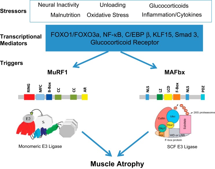
Muscle RING finger 1 (MuRF1) and muscle atrophy F-box (MAFbx)/atrogin-1 were identified more than 10 years ago as two muscle-specific E3 ubiquitin ligases that are increased transcriptionally in skeletal muscle under atrophy-inducing conditions, making them excellent markers of muscle atrophy. In the past 10 years much has been published about MuRF1 and MAFbx with respect to their mRNA expression patterns under atrophy-inducing conditions, their transcriptional regulation, and their putative substrates. However, much remains to be learned about the physiological role of both genes in the regulation of mass and other cellular functions in striated muscle. Although both MuRF1 and MAFbx are enriched in skeletal, cardiac, and smooth muscle, this review will focus on the current understanding of MuRF1 and MAFbx in skeletal muscle, highlighting the critical questions that remain to be answered.

Keywords: atrogenes; muscle RING finger 1; muscle atrophy F-box; muscle sparing; protein quality control; ubiquitin proteasome system.


Figures

Fig. 1.
Fig. 1.
Regulation of muscle RING finger 1 (MuRF1) and muscle atrophy F-box (MAFbx) expression in skeletal muscle. Skeletal muscle atrophy is induced by a number of stressors, as illustrated here. These stressors can lead to the increase in the expression of a number of transcription factors, including the forkhead transcription factors (FOXO1 and FOX03a), NF-κB transcription factors (p65, c-Rel, RelB, p52, and p50), CCAAT/enhancer-binding protein-β (C/EBPβ), kruppel-like factor-15 (KLF-15), and/or activation of the glucocorticoid receptor. These transcriptional mediators can bind to the promoter regions of either the MuRF1 or MAFbx genes, leading to an increase in their expression levels within the muscle. A schematic of the putative domain structure of each protein is shown. MuRF1 contains a RING finger domain (RING), a MuRF1 family conserved domain (MFC), a B-box domain (B-Box), coiled-coil domains (CC), and an acidic tail region (AR). MAFbx contains 2 nuclear localization signals (NLS), a leucine-zipper domain (LZ), a leucine-charged residue-rich domain (LCD), an F-box domain (F-box), and a PDZ domain (PDZ). SCF, Skp1-Cullin1-F-box protein.
Fig. 2.
Fig. 2.
The ubiquitin (Ub)-proteasome Pathway. The process of ubiquitination is controlled by the Ub-activating enzymes (E1), Ub-conjugating enzymes (E2), and Ub-protein ligases (E3). Both MuRF1 and MAFbx are E3 Ub ligases that control the ubiquitination of specific substrates. Ub can be added to a substrate as a single monomer on one (monoubiquitination) or more lysines (multiubiquitination) or as a chain of ubiquitins of variable length on a single lysine (polyubiquitination). The model of ubiquitination can lead to different substrate fates. Polyubiquitination of a substrate with a ubiquitin chain using K11 or K48 linkages generally results in proteasomal degradation, whereas Ub chains using K63 linkages can lead to alterations in signaling or endocytosis. RBX, RING finger protein.
Fig. 3.
Fig. 3.
Phenotype of MuRF1-null [knockout (KO)] mice. The major differences in the response of the MuRF1-KO mice relative to WT mice are highlighted for glucocorticoid treatment, denervation, and aging (18–24 mo old). A common phenotype of all of the models is muscle sparing. Nedd4, neural precursor cell expressed developmentally downregulated protein 4; HDAC4, histone deacetylase 4; ER, endoplasmic reticulum.

References

    1. Adams V, Mangner N, Gasch A, Krohne C, Gielen S, Hirner S, Thierse HJ, Witt CC, Linke A, Schuler G, Labeit S. Induction of MuRF1 is essential for TNF-alpha-induced loss of muscle function in mice. J Mol Biol 384: 48–59, 2008 - PubMed
    1. Allen DL, Bandstra ER, Harrison BC, Thorng S, Stodieck LS, Kostenuik PJ, Morony S, Lacey DL, Hammond TG, Leinwand LL, Argraves WS, Bateman TA, Barth JL. Effects of spaceflight on murine skeletal muscle gene expression. J Appl Physiol 106: 582–595, 2009 - PMC - PubMed
    1. Allen DL, Cleary AS, Lindsay SF, Loh AS, Reed JM. Myostatin expression is increased by food deprivation in a muscle-specific manner and contributes to muscle atrophy during prolonged food deprivation in mice. J Appl Physiol 109: 692–701, 2010 - PubMed
    1. Almon RR, DuBois DC, Yao Z, Hoffman EP, Ghimbovschi S, Jusko WJ. Microarray analysis of the temporal response of skeletal muscle to methylprednisolone: comparative analysis of two dosing regimens. Physiol Genomics 30: 282–299, 2007 - PMC - PubMed
    1. Altun M, Besche HC, Overkleeft HS, Piccirillo R, Edelmann MJ, Kessler BM, Goldberg AL, Ulfhake B. Muscle wasting in aged, sarcopenic rats is associated with enhanced activity of the ubiquitin proteasome pathway. J Biol Chem 285: 39597–39608, 2010 - PMC - PubMed

Publication types

MeSH terms