. 2015 Jan;43 Suppl 1(0 1):13-7.
doi: 10.1007/s00240-014-0702-z. Epub 2014 Aug 6.

Micro-CT imaging of Randall's plaques

Affiliations

Micro-CT imaging of Randall's plaques

James C Williams Jr et al. Urolithiasis. 2015 Jan.

Abstract

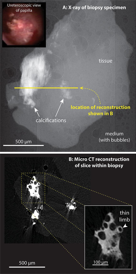
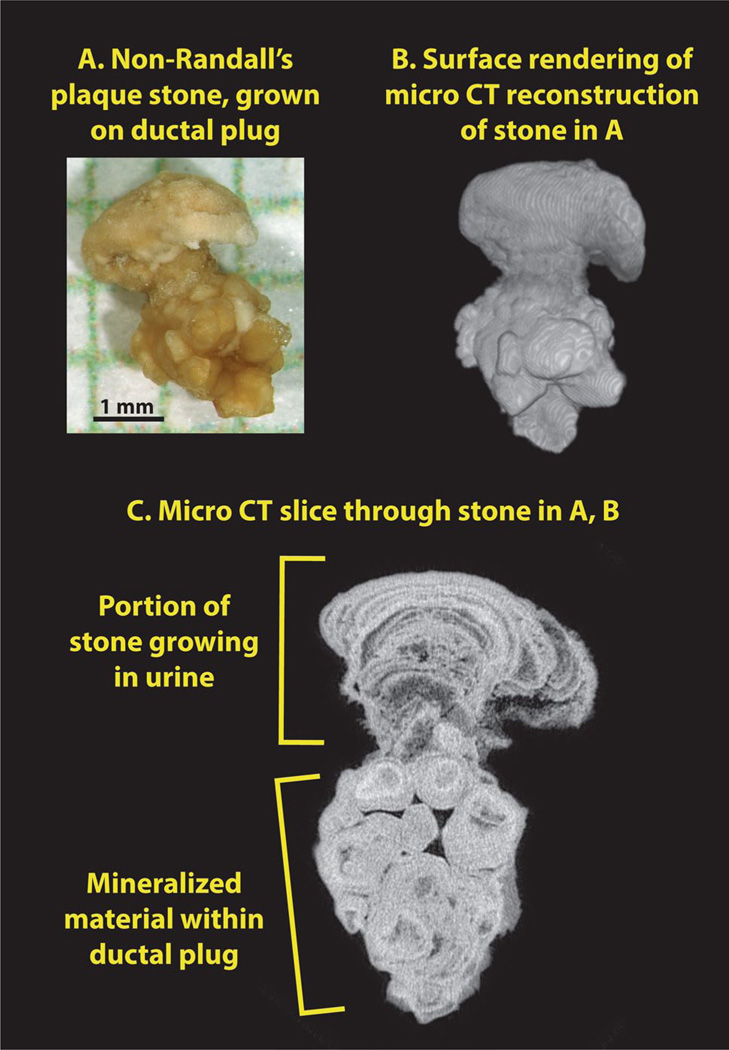
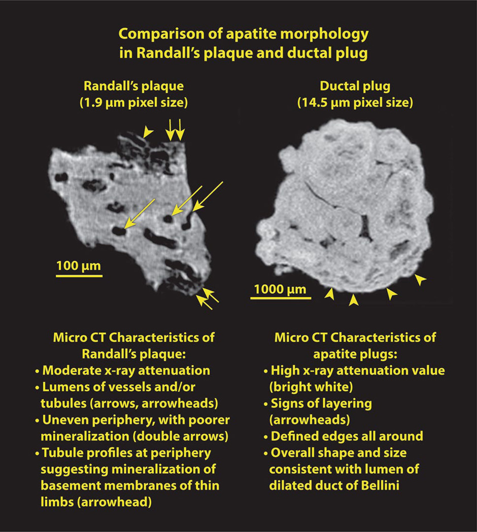
Micro-computed tomographic imaging (micro-CT) provides unprecedented information on stone structure and mineral composition. High-resolution micro-CT even allows visualization of the lumens of tubule and/or vessels within Randall's plaque, on stones or in papillary biopsies, thus giving a non-destructive way to study these sites of stone adhesion. This paper also shows an example of a stone growing on a different anchoring mechanism: a mineral plug within the lumen of a Bellini duct (BD plug). Micro-CT shows striking structural differences between stones that have grown on Randall's plaque and those that have grown on BD plugs. Thus, Randall's plaque can be distinguished by micro-CT, and this non-destructive method shows great promise in helping to elucidate the different mechanisms by which small stones are retained in the kidney during the development of nephrolithiasis.


Conflict of interest statement

Conflict of interest T he authors declare that they have no conflict of interest.

Figures

Fig. 1
Fig. 1
Micro-CT imaging of Randall’s plaque in human papillary biopsy. a Inset ureteroscopic image of papillary surface before biopsy. a X-ray image of biopsy. About 450 such images were taken, with the specimen rotated 0.4° between each image. This image series was used for tomographic reconstruction. b Reconstructed slice through the biopsy, with inset showing higher magnification of a small portion of Randall’s plaque within the tissue
Fig. 2
Fig. 2
Small calcium oxalate monohydrate stone, growing on Randall’s plaque. a Ureteroscopic view of stone (arrowhead) before removal. b Surface rendering of the 3D micro-CT image stack shows a relatively smooth stone surface (top) with some polyhedral calcium oxalate dihydrate (COD) crystals, but a ragged surface for the Randall’s plaque, which was pulled off the papilla tip. c Micro-CT image slice showing stone attached to plaque. Some lumens of tubules and/or vessels within the plaque are marked with arrows
Fig. 3
Fig. 3
Apatite stone growing on end of BD plug. a Photograph of stone, after removal, shown on mm-grid paper. b Surface reconstruction of stone, showing head of stone on top of apatite plug. c Micro-CT image slice, showing the distinct morphologies of the apatite portion that grew in the calyceal urine and the apatite that formed within the BD lumen
Fig. 4
Fig. 4
Randall’s plaque and BD plug, both composed of apatite, shown side by side. See text for details

References

    1. Cleveland RO, McAteer JA, Müller R. Time-lapse non-destructive assessment of shock wave damage to kidney stones in vitro using micro-computed tomography. J Acoust Soc Am. 2001;110:1733–1736. - PubMed
    1. Williams JC, Jr, McAteer JA, Evan AP, Lingeman JE. Micro-computed tomography for analysis of urinary calculi. Urol Res. 2010;38:477–484. - PubMed
    1. Qu M, et al. Dual-energy dual-source CT with additional spectral filtration can improve the differentiation of non-uric acid renal stones: an ex vivo phantom study. Am J Roentgenol. 2011;196:1279–1287. - PMC - PubMed
    1. Evan AP, et al. Randall’s plaque of patients with nephrolithiasis begins in basement membranes of thin loops of Henle. J Clin Invest. 2003;111:607–616. - PMC - PubMed
    1. Evan AP, et al. Mechanism of formation of human calcium oxalate renal stones on Randall’s plaque. Anat Rec. 2007;290:1315–1323. - PubMed

Publication types

MeSH terms

LinkOut - more resources