. 2014 Oct 1;127(Pt 19):4234-45.
doi: 10.1242/jcs.152470. Epub 2014 Aug 5.

Promiscuous methionyl-tRNA synthetase mediates adaptive mistranslation to protect cells against oxidative stress

Affiliations

Promiscuous methionyl-tRNA synthetase mediates adaptive mistranslation to protect cells against oxidative stress

Jin Young Lee et al. J Cell Sci. .

Abstract

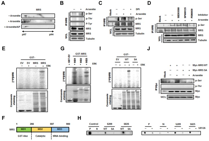
Aminoacyl-tRNA synthetases (ARSs) acylate transfer (t)RNAs with amino acids. Charging tRNAs with the right amino acids is the first step in translation; therefore, the accurate and error-free functioning of ARSs is an essential prerequisite for translational fidelity. A recent study found that methionine (Met) can be incorporated into non-Met residues of proteins through methionylation of non-cognate tRNAs under conditions of oxidative stress. However, it was not understood how this mis-methionylation is achieved. Here, we report that methionyl-tRNA synthetase (MRS) is phosphorylated at Ser209 and Ser825 by extracellular signal-related kinase (ERK1/2) under conditions of stress caused by reactive oxygen species (ROS), and that this phosphorylated MRS shows increased affinity for non-cognate tRNAs with lower affinity for tRNA(Met), leading to an increase in Met residues in cellular proteins. The expression of a mutant MRS containing the substitutions S209D and S825D, mimicking dual phosphorylation, reduced ROS levels and cell death. This controlled inaccuracy of MRS seems to serve as a defense mechanism against ROS-mediated damage at the cost of translational fidelity.

Keywords: Cell protection; ERK; Methionyl-tRNA synthetase; Misacylation; Reactive oxygen species.


Figures

Fig. 1.
Fig. 1.
Determination of ERK-mediated phosphorylation sites in MRS during ROS stress. (A) Lysates from untreated and sodium-arsenite-treated HeLa cells were subjected to 2D-PAGE. The gel was immunoblotted with an anti-MRS antibody. To check ROS-dependent phosphorylation of MRS, lysates from sodium-arsenite-treated cells were incubated with alkaline phosphatase (AP). (B) Lysates prepared as above were immunoprecipitated (IP) with the anti-MRS antibody and immunoblotted with antibodies against phosphoserine (p-Ser), phosphothreonine (p-Thr) and phosphotyrosine (p-Tyr). WCL, whole-cell lysate. (C) Lysates from HeLa cells treated with sodium arsenite with or without DPI (ROS inhibitor) were immunoprecipitated using the anti-MRS antibody. The gel was immunoblotted with antibody against p-Ser. (D) HeLa cells pre-treated with each MAPK inhibitor – SB203580 (p38 MAPK inhibitor), SP600125 (JNK inhibitor) and PD98059 (ERK inhibitor) – were incubated in medium containing sodium arsenite. The lysates were immunoprecipitated with the anti-MRS antibody and immunoblotted with antibody against p-Ser. (E) Purified GST (EV, empty vector) and GST–MRS were subjected to an in vitro kinase reaction by incubating with ERK and [γ-32P]ATP. After staining with Coomassie Brilliant Blue, radioactivity was detected by autoradiography. (F) Schematic representation of functional domains in human MRS. The domains of MRS can be divided into MD1 (GST-like, residues 1–266), MD2 (Catalytic, residues 267–597) and MD3 (tRNA-binding, residues 598–900) fragments. (G) Individual GST-fused domains of MRS were subjected to an in vitro kinase assay with ERK in the presence of [γ-32P]ATP. Phosphorylation signal was detected by autoradiography. (H) HEK293T cells transfected with GFP–ERK were immunoprecipitated with anti-GFP antibody. The immunoprecipitated GFP–ERK was mixed with each biotinylated synthetic peptide (WT, wild type; SA, phosphorylation-inactive form) and [γ-32P]ATP. A consensus sequence phosphorylated by ERK was used as a control (P, positive control; N, phosphorylation-inactive form of positive control) (left). Positive control peptide and peptides containing each S209 and S825 were incubated with [γ-32P]ATP and immunoprecipitated ERK from cells pre-treated with U0126. Autoradioactivity from the peptides was detected after dot blotting (right). (I) To confirm the ERK-dependent phosphorylation sites in MRS, an in vitro kinase assay was performed with wild-type GST–MRS and the GST–MRS-S209A/S825A (SA) mutant as described above. The phosphorylation signal was detected by autoradiography. The relative quantification obtained by densitometric analysis for the phosphorylation signal of wild-type MRS and MRS-SA was 1 and 0.11, respectively. (J) HEK293T cells transfected with wild-type Myc–MRS or the Myc–MRS-SA mutant were treated with sodium arsenite and immunoprecipitated using the anti-Myc antibody. Arsenite-dependent phosphorylation of MRS was detected using the antibody against p-Ser.
Fig. 2.
Fig. 2.
Dually phosphorylated MRS undergoes a conformational change and binds to tRNALys(CUU). (A) Maltose-binding protein (MBP)-tagged wild-type (WT) MRS and the MBP–MRS-SA and SD mutants were purified, and the circular dichroism spectra of these proteins were obtained in the far-UV at two different temperatures (left). The purified proteins were separated by using SDS-PAGE and stained with Coomassie Brilliant Blue (right). (B) Binding affinities of wild-type MRS and the S209D/S825D (SD) mutant to tRNALys(CUU) (upper panel) and tRNAeMet(CAU) (lower panel) were determined by EMSA. Each tRNA probe was incubated with wild-type His–MRS and His–MRS-SD proteins and separated by non-denaturing PAGE.
Fig. 3.
Fig. 3.
Dually phosphorylated MRS induces mismethionylation under ROS stress. (A) HEK293T cells were transfected with wild-type (WT) TagRFP or the TagRFP M67K mutant and the fluorescence of each protein was compared (left). Insets show the same field as in the phase-contrast image. Scale bar: 80 µm. The expression level of wild-type TagRFP and the TagRFP M67K mutant were determined by immunoblotting (right). (B–E) MRS-dependent Met-misincorporation was monitored using the TagRFP M67K mutant, the fluorescence of which disappeared due to the M to K substitution. HEK293T cells co-transfected with Tag-RFP M67K and empty vector (EV) or each type of Myc–MRS (WT, SA or SD mutant) were treated with sodium arsenite. Revival of fluorescence due to Met-misincorporation at the M67K residue position was observed by fluorescence microscopy (×200). Insets show the same field as in the phase-contrast images. Scale bar: 80 µm (B). Expression levels of total MRS, Myc–MRS (wild type, SA or SD mutant) and TagRFP M67K mutant were analyzed by immunoblotting (C). The relative number (D) and the relative fluorescence intensity (E) of red fluorescent cells are presented as bar graphs. Data are represented as the mean±s.d. (n = 3); *P<0.05; **P<0.01; ***P<0.001; a, P-value indicates a significant difference between the arsenite-untreated and -treated groups; b, P-value indicates a significant difference between arsenite-untreated empty vector and SD groups. (F) HEK293T cells transfected with FLAG-VN–AIMP3 were incubated with arsenite, [35S]Met and with or without the ERK inhibitor U0126. The radioactive signal from the immunoprecipitated (IP) FLAG-VN–AIMP3 was detected by autoradiography. WCL, whole-cell lysate. (G) HEK 293T cells co-transfected with FLAG-VN–AIMP3 and empty vector or each type of Myc–MRS (wild type, SA or SD mutant) were incubated with [35S]Met in the presence with arsenite. To see the effect of ERK inhibitor, cells were pre-treated with U0126 1 h before the arsenite treatment. The cell extracts were immunoprecipitated with anti-FLAG antibody. [35S]Met signals from the FLAG-VN–AIMP3 were monitored by autoradiography.
Fig. 4.
Fig. 4.
Met-misincorporated residues in Flag-VN–AIMP3 and their location in the AIMP3 and Venus structures. (A,B) The Met-exchanged residues identified by mass spectrometry analysis are depicted in the AIMP3 (pdb2uz8) (A) and the full Venus (pdb3t6h) (B) structures. Residues only identified from HEK293T cells expressing wild-type (WT) MRS (A,B, upper images) and MRS-SD (A,B, middle images) are depicted in cyan and red, respectively. Common residues identified from both cells are represented in orange. The original Met residue in AIMP3 is depicted in green. Mismethionylated residues on AIMP3 in MRS-SD-overexpressing conditions are also represented on the MRS–AIMP3 complex structure (pdb4bl7) (A, lower images). The GST-domain of MRS is shown in light green. Each N-terminus of Venus (VN) and deleted C-terminus of Venus (VC) in the full Venus protein structure is shown in green and red, respectively, for convenience (B, lower panel).
Fig. 5.
Fig. 5.
Dually phosphorylated MRS reduces intracellular ROS levels and promotes cell survival under ROS stress. (A) HEK293T cells were transfected with empty vector (EV) or Myc-tagged wild-type (WT) MRS, or the SA or SD mutants, and incubated with arsenite. ROS levels were detected by the DCFH-DA assay. Insets show the same field as in the phase-contrast images. Scale bar: 200 µm. (B) Bax and Bcl-2 levels were detected with their specific antibodies under the same conditions as shown in A. Exogenous (Exo) and endogenous (Endo) MRS were separated and detected using the anti-MRS antibody to show the expression level. (C) MRS level in HEK293T cells was reduced by treatment with MRS-specific siRNA (si-MRS) for 72 h. The effect of MRS expression on cell viability under ROS stress was determined by the MTT assay. (D,E) The effect of MRS proteins on cell viability under ROS stress was determined by the MTT assay. MRS proteins were transiently expressed in HEK293T cells (D) and stably expressed in HeLa cells (E). In C–E: a, P-value indicates a significant difference between the arsenite-untreated and -treated groups; b, P-value indicates a significant difference between arsenite-treated si-control and si-MRS groups. (F) The growth curves of MRS-expressing stably transfected HeLa cells were monitored in the presence and absence of ROS stress. (G) ROS-dependent apoptosis was determined in MRS-expressing stably transfected HeLa cells using the TUNEL assay. Cells incubated with or without arsenite for 72 h were fixed and stained with 4′,6-diamidino-2-phenylindole (DAPI; blue) and fluorescein-labeled dUTP. Green fluorescence indicates TUNEL-positive cells. Scale bar: 200 µm (upper panel). The number of TUNEL-positive cells was normalized to that of DAPI-positive cells, and the quantitative analysis is shown (lower panel). (H) HeLa cells expressing wild-type MRS were subjected to TUNEL assay as described for G with or without U0126 pre-treatment, and the images (upper panel) and the quantitative analysis (lower panel) for positive cells are shown. Scale bar: 200 µm. All quantitative data show the mean±s.d. (n = 3);*P<0.05; **P<0.01; ***P<0.001.
Fig. 6.
Fig. 6.
Schematic model for the protective role of MRS under ROS stress. Upon ROS stress, ERK is activated and phosphorylates MRS at the Ser209 and Ser825 residues. Phosphorylated MRS enhances the mischarging of Met on non-methionyl tRNAs, such as tRNALys. Met carried by non-cognate tRNAs is incorporated into growing polypeptides during translation and used as a ROS scavenger, protecting cells from oxidative damage and apoptosis.

References

    1. Arici T., Dikbas S., Altunbasak Y. (2009). A histogram modification framework and its application for image contrast enhancement. IEEE Trans. Image Process. 18, 1921–1935 10.1109/TIP.2009.2021548 - DOI - PubMed
    1. Birben E., Sahiner U. M., Sackesen C., Erzurum S., Kalayci O. (2012). Oxidative stress and antioxidant defense. World Allergy Organ. J. 5, 9–19 10.1097/WOX.0b013e3182439613 - DOI - PMC - PubMed
    1. Chen Y. C., Lin-Shiau S. Y., Lin J. K. (1998). Involvement of reactive oxygen species and caspase 3 activation in arsenite-induced apoptosis. J. Cell. Physiol. 177, 324–333 10.1002/(SICI)1097-4652(199811)177:2<324::AID-JCP14>3.0.CO;2-9 - DOI - PubMed
    1. Englisch S., Englisch U., von der Haar F., Cramer F. (1986). The proofreading of hydroxy analogues of leucine and isoleucine by leucyl-tRNA synthetases from E. coli and yeast. Nucleic Acids Res. 14, 7529–7538 10.1093/nar/14.19.7529 - DOI - PMC - PubMed
    1. Fersht A. R., Dingwall C. (1979). An editing mechanism for the methionyl-tRNA synthetase in the selection of amino acids in protein synthesis. Biochemistry 18, 1250–1256 10.1021/bi00574a021 - DOI - PubMed

Publication types

LinkOut - more resources