

Comment
doi: 10.7554/eLife.03811.
The spinal cord is never at rest
Affiliations
- PMID: 25097250
- PMCID: PMC4120650
- DOI: 10.7554/eLife.03811
Item in Clipboard
Comment
The spinal cord is never at rest
Elife.
.
Abstract
Even when we are at rest, our spinal cords show spontaneous, yet well organised, fluctuations of activity that might reflect sensory and motor networks.
Keywords: 7 Tesla; fMRI; functional connectivity; resting state; spinal cord.
Copyright © 2014, Eippert and Tracey.
Conflict of interest statement
Figures
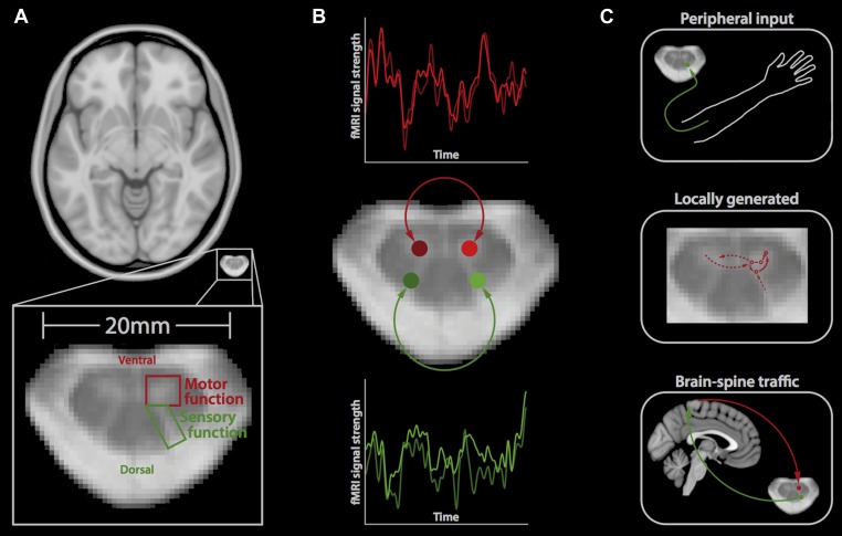
(A) Horizontal section of a brain (top) and a spinal cord (middle, bottom); the small size of the spinal cord makes it difficult to image neuronal activity. The spinal cord contains two ventral horns (one outlined in red) that are involved in motor function, and two dorsal horns (one outlined in green) that are involved in sensory function. (B) Barry et al. measured the correlation between spontaneous fluctuations in the fMRI signal in the ventral horns (red traces; top) and the dorsal horns (green traces; bottom). This revealed that the ventral horns show a positive correlation with each other, as do the dorsal horns. However, there is no significant correlation between ventral and dorsal horns. This suggests that at rest, the spinal cord is intrinsically organised into two separate networks, corresponding to motor and sensory functions. (C) Possible mechanisms that could explain the spontaneous activity in the spinal cord include input from the peripheral nervous system (top), locally generated rhythms from the interneurons within spinal networks (middle), and ongoing communication between the brain and spinal cord (bottom).
Comment on
-
Resting state functional connectivity in the human spinal cord.Elife. 2014 Aug 5;3:e02812. doi: 10.7554/eLife.02812. Elife. 2014. PMID: 25097248 Free PMC article.
References
Publication types
MeSH terms
LinkOut - more resources
Full Text Sources
Other Literature Sources
