

Radiosensitivity of human ovarian carcinoma and melanoma cells to γ-rays and protons
- PMID: 25097591
- PMCID: PMC4107263
- DOI: 10.5114/aoms.2014.43751
Radiosensitivity of human ovarian carcinoma and melanoma cells to γ-rays and protons
Abstract
Introduction: Proton radiation offers physical advantages over conventional radiation. Radiosensitivity of human 59M ovarian cancer and HTB140 melanoma cells was investigated after exposure to γ-rays and protons.
Material and methods: Irradiations were performed in the middle of a 62 MeV therapeutic proton spread out Bragg peak with doses ranging from 2 to 16 Gy. The mean energy of protons was 34.88 ±2.15 MeV, corresponding to the linear energy transfer of 4.7 ±0.2 keV/µm. Irradiations with γ-rays were performed using the same doses. Viability, proliferation and survival were assessed 7 days after both types of irradiation while analyses of cell cycle and apoptosis were performed 48 h after irradiation.
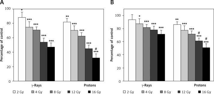
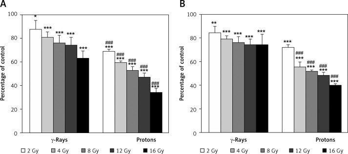
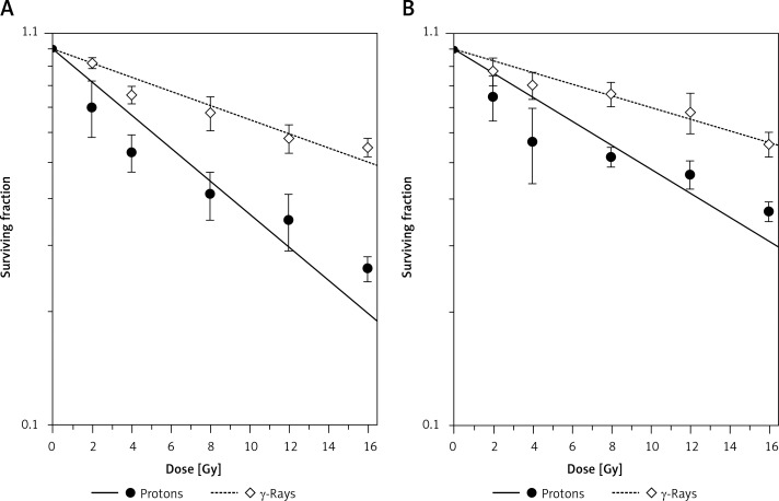
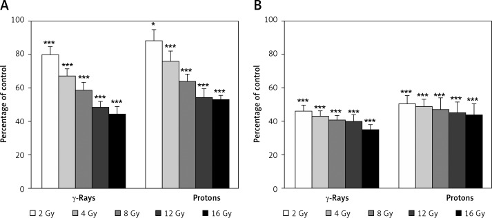
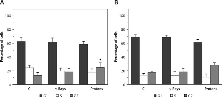
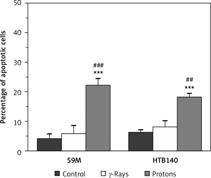
Results: Results showed that γ-rays and protons reduced the number of viable cells for both cell lines, with stronger inactivation achieved after irradiation with protons. Surviving fractions for 59M were 0.91 ±0.01 for γ-rays and 0.81 ±0.01 for protons, while those for HTB140 cells were 0.93 ±0.01 for γ-rays and 0.86 ±0.01 for protons. Relative biological effectiveness of protons, being 2.47 ±0.22 for 59M and 2.08 ±0.36 for HTB140, indicated that protons provoked better cell elimination than γ-rays. After proton irradiation proliferation capacity of the two cell lines was slightly higher as compared to γ-rays. Proliferation was higher for 59M than for HTB140 cells after both types of irradiation. Induction of apoptosis and G2 arrest detected after proton irradiation were more prominent in 59M cells.
Conclusions: The obtained results suggest that protons exert better antitumour effects on ovarian carcinoma and melanoma cells than γ-rays. The dissimilar response of these cells to radiation is related to their different features.
Keywords: apoptosis; cell cycle; melanoma; ovarian carcinoma; protons; γ-rays.
Figures
References
-
- Gordon AT, McMillan TJ. A role for molecular radiobiology in radiotherapy? Clin Oncol (R Coll Radiol) 1997;9:70–8. - PubMed
-
- Steel GG. Introduction: The significance of radiobiology for radiotherapy. In: Steel GG, editor. Basic clinical radiobiology. London: Edward Arnold; 1993. pp. 1–7.
-
- Petrović I, Ristić-Fira A, Todorović D, Valastro L, Cirrone P, Cuttone G. Radiobiological analysis of human melanoma cells on the 62 MeV CATANA proton beam. Int J Radiat Biol. 2006;82:251–65. - PubMed
-
- Soengas MS, Lowe SW. Apoptosis and melanoma chemoresistance. Oncogene. 2003;22:3138–51. - PubMed
LinkOut - more resources
Full Text Sources
Other Literature Sources