Review
. 2014 Sep;14(9):598-610.
doi: 10.1038/nrc3792. Epub 2014 Aug 7.

FAK in cancer: mechanistic findings and clinical applications

Affiliations
Review

FAK in cancer: mechanistic findings and clinical applications

Florian J Sulzmaier et al. Nat Rev Cancer. 2014 Sep.

Abstract

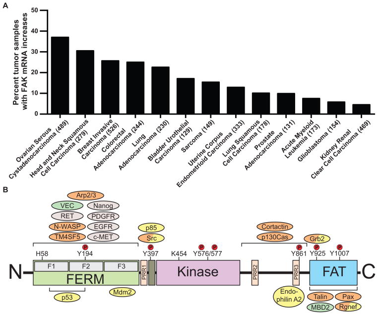
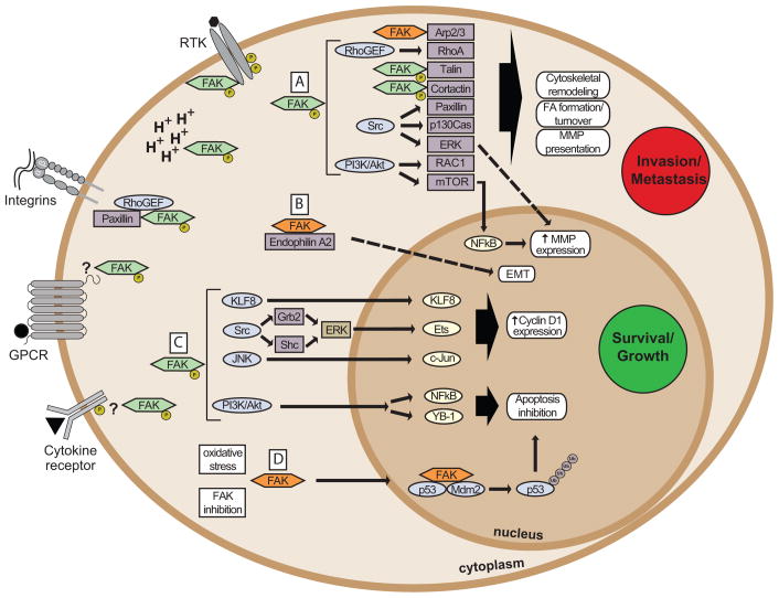
Focal adhesion kinase (FAK) is a cytoplasmic protein tyrosine kinase that is overexpressed and activated in several advanced-stage solid cancers. FAK promotes tumour progression and metastasis through effects on cancer cells, as well as stromal cells of the tumour microenvironment. The kinase-dependent and kinase-independent functions of FAK control cell movement, invasion, survival, gene expression and cancer stem cell self-renewal. Small molecule FAK inhibitors decrease tumour growth and metastasis in several preclinical models and have initial clinical activity in patients with limited adverse events. In this Review, we discuss FAK signalling effects on both tumour and stromal cell biology that provide rationale and support for future therapeutic opportunities.


Conflict of interest statement

Competing interests statement

The authors declare no competing financial interests.

Figures

Figure 1
Figure 1. FAK expression in cancer and FAK domain structure
(A) Percent of tumor samples with elevated focal adhesion kinase (FAK) mRNA. The Cancer Genome Atlas was quiered using the cBioPortal (www.cbioportal.org). Search criteria included mRNA expression data (Z-scores for all genes) and tumor datasets with mRNA data. Numbers of tumors analyzed (n) is shown on the X axis. (B) FAK consists of a central kinase domain flanked by a protein4.1-ezrin-radixin-moesin (FERM) homology domain on the N-terminal side and a C-terminal focal adhesion targeting (FAT) domain. Both terminal domains are separated from the kinase domain by a linker region containing proline-rich regions (PRR). Important tyrosine (Y) phosphorylation (P) sites are indicated; Y397, K454 and H58 play crucial roles in FAK activation. FAK binding partners are shown at their interaction sites within FAK. Binding of these proteins affects outcomes like cell motility (orange), cell survival (yellow) or both functions (orange/yellow). Roles involving FAK activation are shown in grey, important contributions to the tumor environment in green.
Figure 2
Figure 2. FAK connections to tumor growth and metastasis
FAK drives cancer growth and metastasis through kinase-dependent (green) or -independent (orange) functions. FAK is activated by receptor tyrosine kinases (RTK), intracellular pH changes (H+), integrins, G-protein coupled receptors (GPCR) and cytokine receptors. The exact mechanisms are not always clear (indicated by ‘?’). Oxidative stress and FAK catalytic inhibition increase FAK nuclear localization. (A) Active FAK increases cell motility through effects on Arp2/3, Rho guanine nucleotide exchange factors (RhoGEF), talin or cortactin, and Src- or PI3-kinase (PI3K) mediated signaling. This drives cytoskeletal remodeling, focal adhesion (FA) formation and turnover, and expression and cell-surface presentation of matrix metalloproteinases (MMPs), enhancing cell invasion and tumor metastasis. (B) Kinase-independent scaffolding of endophilin A2 induces the expression of endothelial-mesenchymal transition (EMT) markers. FAK affects survival and proliferation through kinase-dependent and –independent roles to promote tumor growth. (C) FAK induces cell cycle progression through cyclin D1 involving Krüppel-like factor 8 (KLF8), Src–ERK, or JNK signaling. Signaling through PI3K/Akt mediates inhibition of apoptosis through transcriptional effects by NFkB or YB-1. (D) Nuclear FAK acts as a scaffold for p53 and Mdm2 in a kinase-independent manner, increasing p53 poly-ubiquitination (Ub) and degradation, thereby promoting cell survival.
Figure 3
Figure 3. Regulation of vascular permeability and extravasation processes by endothelial cell FAK
Tumor cells, via the secretion of growth factors and the activation EC-specific receptors (1) induce conformational FAK activation through multiple mechanisms. (2) FAK signaling promotes ERK/MAPK signaling cascade activation (3) leading to GATA-4-dependent transcriptional expression of VCAM-1 (4), a surface protein that can facilitate immune cell adhesion to ECs (5). EC FAK also promotes E-selectin expression (6) favoring tumor cell adhesion to ECs (7) and the lodging of metastatic cancer cells within sites of vascular hyper-permeability. FAK activation can occur downstream of integrin receptor binding to matrix proteins consisting of a FAK-Src multi-protein complex (8). In response to VEGF signals, FAK promotes the localization of Src to adherens junctions; key sites that maintain vascular barrier integrity (9). FAK binding via FERM domain to the VE-cadherin (VEC) cytoplasmic tail, and FAK-dependent pY658 VEC and pY142 β-catenin phosphorylation (10) promotes VEC/β-catenin dissociation and VEC internalization/degradation. Loss of VEC from the cell surface leads to increased vascular permeability (11), allows for tumor cell transmigration across EC barriers, and leads to increased tumor cell metastasis (12), .
Figure 4
Figure 4. Tumor microenvironmental impact of FAK signals
FAK is an important regulator of EC, neutrophil, platelet, macrophage and fibroblast signaling in the tumor microenvironment leading to the increase (green boxes) or decrease (red boxes) of stromal cell functions. In ECs, FAK inhibits apoptosis and increases proliferation. EC FAK also contributes to the formation of abnormal vasculature via the increase of cell migration, survival, and vascular permeability (VP). Moreover, as described in detail in Figure 3, EC FAK is a key regulator of vascular permeability and tumor intra/extravasation leading to metastasis. FAK stimulates macrophage and fibroblast migration. FAK promotes the differentiation (dotted arrow) of macrophages. This occurs in stimulus-specific fashion where FAK activation either promotes or reverses (red/green box) fibroblast differentiation into CAF. For both macrophages and fibroblasts, FAK activity positively impacts cell recruitment to the tumor site. FAK promotes spreading, adhesion, and survival of stromal cells; with concomitant regulation ECM synthesis/remodeling to promote tumor progression. References are available in the corresponding text. VP: vascular permeability, TC: tumor cell, EC: endothelial cell, TAM: tumor-associated macrophage and CAF: cancer-associated fibroblast.

References

    1. Parsons JT. Focal adhesion kinase: the first ten years. J Cell Sci. 2003;116:1409–16. - PubMed
    1. Schaller MD. Cellular functions of FAK kinases: insight into molecular mechanisms and novel functions. J Cell Sci. 2010;123:1007–13. - PubMed
    1. Zhao J, Guan JL. Signal transduction by focal adhesion kinase in cancer. Cancer Metastasis Rev. 2009;28:35–49. - PubMed
    1. Goode EL, et al. A genome-wide association study identifies susceptibility loci for ovarian cancer at 2q31 and 8q24. Nat Genet. 2010;42:874–9. - PMC - PubMed
    1. Cancer Genome Atlas Research N. Integrated genomic analyses of ovarian carcinoma. Nature. 2011;474:609–15. - PMC - PubMed

Publication types

MeSH terms