. 2014 Sep 15;193(6):2961-70.
doi: 10.4049/jimmunol.1400728. Epub 2014 Aug 6.

The immunotherapeutic role of regulatory T cells in Leishmania (Viannia) panamensis infection

Affiliations

The immunotherapeutic role of regulatory T cells in Leishmania (Viannia) panamensis infection

Allison Ehrlich et al. J Immunol. .

Abstract

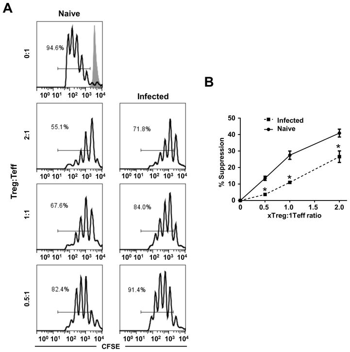
Leishmania (Viannia) parasites are etiological agents of cutaneous leishmaniasis in the New World. Infection is characterized by a mixed Th1/Th2 inflammatory response, which contributes to disease pathology. However, the role of regulatory T cells (Tregs) in Leishmania (Viannia) disease pathogenesis is unclear. Using the mouse model of chronic L. (V.) panamensis infection, we examined the hypothesis that Treg functionality contributes to control of pathogenesis. Upon infection, Tregs (CD4(+)Foxp3(+)) presented with a dysregulated phenotype, in that they produced IFN-γ, expressed Tbet, and had a reduced ability to suppress T cell proliferation in vitro. Targeted ablation of Tregs resulted in enlarged lesions, increased parasite load, and enhanced production of IL-17 and IFN-γ, with no change in IL-10 and IL-13 levels. This indicated that an increased inflammatory response was commensurate with disease exacerbation and that the remaining impaired Tregs were important in regulation of disease pathology. Conversely, adoptive transfer of Tregs from naive mice halted disease progression, lowered parasite burden, and reduced cytokine production (IL-10, IL-13, IL-17, IFN-γ). Because Tregs appeared to be important for controlling infection, we hypothesized that their expansion could be used as an immunotherapeutic treatment approach. As a proof of principle, chronically infected mice were treated with rIL-2/anti-IL-2 Ab complex to expand Tregs. Treatment transitorily increased the numbers and percentage of Tregs (draining lymph node, spleen), which resulted in reduced cytokine responses, ameliorated lesions, and reduced parasite load (10(5)-fold). Thus, immunotherapy targeting Tregs could provide an alternate treatment strategy for leishmaniasis caused by Leishmania (Viannia) parasites.


Figures

Figure 1
Figure 1. Characterization of T regulatory cells in the draining lymph node during L. (V.) panamensis infection
BALB/c mice were infected with 5×10⁴ parasites in the top of the rear foot. At various times post-infection, LN cell populations were removed and T regulatory cell markers were evaluated; LN cells from naïve mice were used as controls. (A) Cells from naïve and chronically infected mice (>12 weeks post-infection) were gated on CD4+Foxp3+ cells and analyzed for expression of CTLA-4, CD25, CD39, and CD73 (solid line, naïve; dashed, chronic infection; shaded, isotype control). (B) SLA-stimulated draining lymph node cells were gated on CD4+ cells and examined at the indicated times post-infection. Tbet and FoxP3 expression are shown. (C) Lymph node cells from chronically infected mice were directly cultured ex vivo with PMA/ionomycin and examined for Foxp3 and IFN-γ. Cells were gated on CD4+Foxp3+ cells. Experiments were independently repeated at least 2 times.*, p≤0.05.
Figure 2
Figure 2. Tregs from L. (V.) panamensis infected mice have reduced suppressive capacity
(A) CFSE labeled CD4+CD25− cells (Teff) from naïve mice were cocultured with irradiated APCs, αCD3, and CD4+CD25+ cells (Treg) from chronically infected or naïve mice at the indicated ratios. Unstimulated CFSE labeled CD4+CD25− cells (shaded line) are shown; CFSE levels are plotted and % proliferated cell indicated. (B) Suppression was calculated using the following equation, (%proliferationTeff − %proliferationTreg+Teff)/%proliferationTeff. Samples were analyzed in triplicate and data are representative of two independent experiments. *, p≤0.05.
Figure 3
Figure 3. Inhibition of IDO exacerbates disease
Mice were infected 5×10⁴ parasites then treated with 1-methyl tryptophan (2mg/ml) ad libitum in their drinking water throughout the course of infection. (A) Lesion size was evaluated throughout the course of infection. At the termination of the experiment (6 weeks) (B) parasite load in the foot was determined using the limiting dilution method and (C) draining lymph node cells were stimulated with SLA for 72 hours. Supernatants were collected and analyzed in duplicate by ELISA. Data represent two independent experiments. *, p≤0.05.
Figure 4
Figure 4. Depletion of T regulatory cells in DEREG mice results in disease exacerbation
DEREG (DTR and eGFP under Foxp3 locus) mice were utilized to specifically deplete Foxp3+ cells. Three weeks post infection (after lesions developed), mice were given 0.5ug diphtheria toxin, i.p. (A) Lesion size was monitored over the course of infection and (B) parasite burden was calculated as described above. (C) At the termination of the experiment, the draining lymph node cells were stimulated with SLA for 72 hours and supernatants were collected and analyzed in duplicate by ELISA. Data represent three independent experiments. *, p≤0.05.
Figure 5
Figure 5. Transfer of regulatory T cells reduces leishmaniasis severity
CD4+CD25+ and CD4+CD25− cells were isolated from the spleens of naïve mice, as described in Materials and Methods section. Three weeks post-infection, 3×10⁵cells (CD4+CD25+ or CD4+CD25−) were injected intralesionally. (A) Lesions were measured throughout the course of infection and (B) parasite burden was analyzed as described above, at the termination of the experiment. (C) Draining lymph node cells were stimulated with SLA for 72 hours. Supernatants were collected and analyzed in duplicate by ELISA. Data represent three independent experiments. *, p≤0.05.
Figure 6
Figure 6. IL-2 antibody complex treatment expands regulatory T cells in vivo
Following the development of lesions (3–4 weeks post-infection), mice were given IL-2/anti-IL-2 in complex as described in Materials and Method section. (A) Representative FACS analysis (gated on CD4+ cells) comparing CD4+Foxp3+ cell populations between control and IL-2 complex treated mice two days post-treatment. (B, C) At two (left) and eight (right) days post-treatment, draining lymph node and spleens were excised and (B) cell numbers and (C) cell populations (CD4+, CD8+ and CD4+FoxP3+) were assessed. (D) Draining lymph node cells from control and treated mice (2 days post-treatment) were gated on CD4+Foxp3+ cells and analyzed for expression of CTLA-4, CD25, CD39, and CD73.
Figure 7
Figure 7. In vivo induction of regulatory T cells improves immunopathology
Following the development of lesions (3–4 weeks post-infection), mice were given IL-2/anti-IL-2 in complex as described in Materials and Method section. (A) Lesions were measured throughout the course of infection and (B) parasite burden was analyzed at the termination of the experiment. (C) Two days following the final treatment and (D) at the termination of the experiment, lymph node cells (C, D) and splenocytes (C) were stimulated with SLA for 72 hours. Supernatants were collected and analyzed in duplicate by ELISA. Data represent two independent experiments *, p≤0.05.

References

    1. Banuls AL, Hide M, Prugnolle F. Leishmania and the leishmaniases: a parasite genetic update and advances in taxonomy, epidemiology and pathogenicity in humans. Adv Parasitol. 2007;64:1–109. - PubMed
    1. Lipoldova M, Demant P. Genetic susceptibility to infectious disease: lessons from mouse models of leishmaniasis. Nat Rev Genet. 2006;7:294–305. - PubMed
    1. Varela MR, Munoz DL, Robledo SM, Kolli BK, Dutta S, Chang KP, Muskus C. Leishmania (Viannia) panamensis: an in vitro assay using the expression of GFP for screening of antileishmanial drug. Exp Parasitol. 2009;122:134–139. - PMC - PubMed
    1. Osorio LE, Castillo CM, Ochoa MT. Mucosal leishmaniasis due to Leishmania (Viannia) panamensis in Colombia: clinical characteristics. Am J Trop Med Hyg. 1998;59:49–52. - PubMed
    1. McMahon-Pratt D, Alexander J. Does the Leishmania major paradigm of pathogenesis and protection hold for New World cutaneous leishmaniases or the visceral disease? Immunol Rev. 2004;201:206–224. - PubMed

Publication types

MeSH terms