. 2013 Mar-Apr;15(2):107-18.
doi: 10.1111/ede.12021.

Manipulation of Fgf and Bmp signaling in teleost fishes suggests potential pathways for the evolutionary origin of multicuspid teeth

Affiliations

Manipulation of Fgf and Bmp signaling in teleost fishes suggests potential pathways for the evolutionary origin of multicuspid teeth

William R Jackman et al. Evol Dev. 2013 Mar-Apr.

Abstract

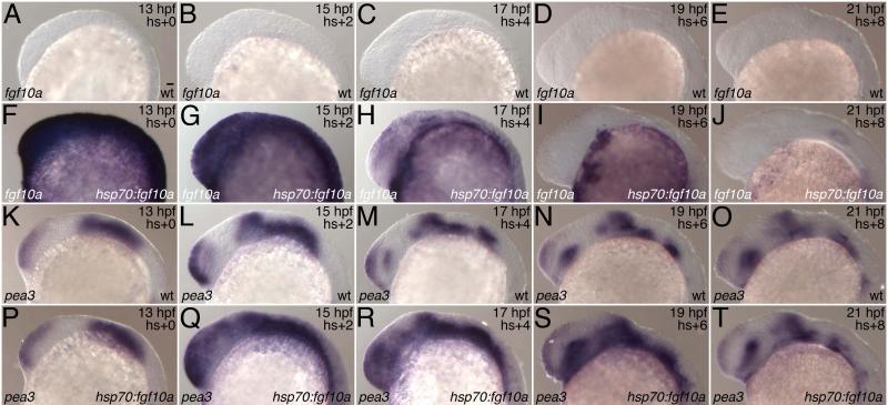
Teeth with two or more cusps have arisen independently from an ancestral unicuspid condition in a variety of vertebrate lineages, including sharks, teleost fishes, amphibians, lizards, and mammals. One potential explanation for the repeated origins of multicuspid teeth is the existence of multiple adaptive pathways leading to them, as suggested by their different uses in these lineages. Another is that the addition of cusps required only minor changes in genetic pathways regulating tooth development. Here we provide support for the latter hypothesis by demonstrating that manipulation of the levels of Fibroblast growth factor (Fgf) or Bone morphogenetic protein (Bmp) signaling produces bicuspid teeth in the zebrafish (Danio rerio), a species lacking multicuspid teeth in its ancestry. The generality of these results for teleosts is suggested by the conversion of unicuspid pharyngeal teeth into bicuspid teeth by similar manipulations of the Mexican Tetra (Astyanax mexicanus). That these manipulations also produced supernumerary teeth in both species supports previous suggestions of similarities in the molecular control of tooth and cusp number. We conclude that despite their apparent complexity, the evolutionary origin of multicuspid teeth is positively constrained, likely requiring only slight modifications of a pre-existing mechanism for patterning the number and spacing of individual teeth.


Figures

Fig. 1
Fig. 1
Evolution of tooth shape in ray-finned fishes (Actinopterygii). The presence of unicuspid teeth is indicated by white shading and of multicuspid teeth by black shading; branches with both colors indicate presence of both character states. Note that unicuspid is the ancestral state for actinopterygian tooth shape. Representative species illustrated above the tree are from left to right Erpetoichthys calabaricus (Reedfish), Pantodon buchholzi (Freshwater Butterflyfish), Gnathonemus petersii (Elephantnose Fish), Danio rerio (Zebrafish), Astyanax mexicanus (Mexican Tetra), Mugil cephalus (Striped Mullet), Limia nigrofasciata (Blackbarred Limia), Xenotoca eiseni (Redtail Splitfin), Lepomis macrochirus (Bluegill), and Maylandia estherae (Red Zebra). Illustrated teeth are premaxillary (upper oral – E. calabaricus, G. petersii, A. mexicanus, M. cephalus, X. eiseni, L. macrochirus, M. estherae), maxillary (upper oral – P. buchholzi), dentary (lower oral – L. nigrofasciata), or fifth ceratobranchial (lower pharyngeal – D. rerio). The phylogeny and composition of orders follow Nelson (2006). Mapping tooth shape on the molecular phylogeny of Near et al. (2012) results in a similar conclusion of multiple origins of multicuspid teeth in ray-finned fishes (supporting information Fig. S1; not shown).
Fig. 2
Fig. 2
Fgf10 overexpression produces supernumerary and bicuspid teeth in the zebrafish. (A) Alizarin-stained zebrafish showing location of fifth ceratobranchial teeth (arrow) in the pharynx. (B-D) Sequence of tooth appearance in wild type zebrafish revealed by alizarin staining. Designations of individual teeth (e.g. 4V1) follow Laurenti et al. (2004) and Van der heyden and Huysseune (2000). (E-P) Dentition of zebrafish overexpressing Fgf10. Teeth are designated as in (B-D), with supernumerary teeth indicated by “S”, two separate homologs of a single wild type tooth by “a” and “b”, and bicuspid teeth by “a-b”, or “4V1-4V1” according to their hypothesized origin. Fish in (E-M) are from transgenic line Tg(hsp70l:fgf10a-GFP)cs2, those in (N, P) were injected with a construct for overexpressing an Fgf10a-Egfp fusion protein and that in (O) was injected with a similar construct for Fgf10b. White rectangles indicate portions of an image captured at a different focal plane. Scale bars = 25 μm. c5, fifth ceratobranchial bone; cl, cleithrum; le, lens; nc, notochord; op, operculum; ot, otolith.
Fig. 3
Fig. 3
Overexpression of multiple Fgf ligands produces supernumerary or bicuspid teeth in the zebrafish. (A) Bicuspid tooth (a-b) induced by implantation of a bead soaked in human Fgf10. Position of bead indicated by asterisk. (B) Phylogenetic tree of Fgf ligands (modified from Itoh and Ornitz 2011) with those found to induce supernumerary or bicuspid teeth upon overexpression indicated by a “+”and those found not to indicated by a “−“. Numbers of injected fish with supernumerary or bicuspid teeth as a fraction of the total injected for ligands other than fgf10a are fgf1 (0/74), fgf3 (2/18), fgf4 (2/58), fgf8a (0/84), fgf10b (3/51), and fgf16 (1/30). (C-D) Supernumerary teeth induced by overexpression of fgf3 and fgf4. Tooth homology in (A, C-D) indicated as in Fig. 2. Scale bar = 25 μm.
Fig 4
Fig 4
Expression of dental markers in wild type (wt) and Fgf10-overexpressing (hsp70:fgf10a) zebrafish. Dorsal views of pitx2 expression (A-C) and ventral views of dlx2b (D-F) and fgf4 (G-I) expression in heat-shocked transgenic line Tg(hsp70l:fgf10a-GFP)cs2 and wild type siblings. Arrows indicate tooth-competent epithelium (A-C) or tooth germs (D-I) on left side of fish. Arrowheads indicate ectopic posterior expression and double arrows two tooth germs appearing in the place of a single germ in the wild type. Scale bars = 25 μm.
Fig. 5
Fig. 5
Time course of expression of fgf10a and its target pea3 following heat shock in transgenic line Tg(hsp70l:fgf10a-GFP)cs2. Expression of fgf10a (A-J) and pea3 (K-T) was determined by in situ hybridization in transgenics (hsp70:fgf10a) and their wild type siblings (wt). All embryos were heat-shocked at 40 °C for 1 hr starting at 12 hpf and fixed at the age indicated in the upper right. Note that fgf10a expression is strongly induced by the end of the heat shock and has largely faded within 8 hr post-heat shock. pea3 expression is strongly induced by 2 hr post-heat shock and has returned to wild type levels by 8 hr post-heat shock. Lateral views with anterior to the left. Scale bar = 25 μm.
Fig. 6
Fig. 6
Supernumerary and bicuspid teeth induced by inhibition of Bmp signaling in the zebrafish. (A) GFP overexpression results in wild type dentition. (B-C). Noggin1 overexpression results in bicuspid (a-b) or supernumerary (a, b) teeth. (D-F) Inhibition of Bmp signaling synergizes with Fgf10 overexpression in the production of supernumerary and multicuspid teeth. Transgenic fish from the line Tg(hsp70l:dnBmpr-GFP)w30 (capable of expressing a dominant negative version of a Bmp receptor) (Pyati et al. 2005) were injected with a construct for overexpressing an Fgf10a-Egfp fusion protein and heat shocked (D-E). Arrows indicate a supernumerary row of teeth in (D) and a tricuspid tooth in (E). (F) Wild type fish were injected with a construct for overexpressing an Fgf10a-Egfp fusion protein, heat shocked and treated with the Bmp inhibitor dorsomorphin. Tooth homology indicated as in Fig. 2. Scale bars = 25 μm. nc, notochord; ot, otolith.
Fig 7
Fig 7
Supernumerary and bicuspid teeth produced in the pharyngeal dentition of Astyanax mexicanus by overexpression of Fgf10 or inhibition of Bmp signaling. (A) Dorsolateral view of upper pharyngeal toothplate with the order of appearance of each tooth indicated (as determined from examination of a developmental series not shown). (B-C) Dorsal views of supernumerary (a, b) and bicuspid (a-b) teeth induced by injection of a construct for overexpressing an Fgf10a-Egfp fusion protein followed by heat shock. Fifth ceratobranchial teeth (designated by “C”) are visible in (B) in addition to teeth of the upper pharyngeal toothplate. (D-F) Dorsolateral views of supernumerary (a, b) and bicuspid (a-b) upper pharyngeal teeth induced by treatment with dorsomorphin. The only abnormal phenotype in (D) is an apparent delay in tooth initiation (also found in E-F), as compared with wild type (A). Tooth homology in (B-F) determined by comparison with (A). Dentition of left and right side visible in (B-C); that of a single side in (A, D-F). Scale bars = 25 μm.

References

    1. Alexander RM. Adaptation in the skulls and cranial muscles of South American characinoid fish. J. Linn. Soc. Lond. Zool. 1964;45:169–190.
    1. Andreucci RD, Britski HA, Carneiro J. Structure and evolution of tetraodontoid teeth: an autoradiographic study (Pisces, Tetraodontiformes). J. Morphol. 1982;171:283–292. - PubMed
    1. Bolt JR. Lissamphibian origins. In: Schultze H-P, Trueb L, editors. Origins of the Higher Groups of Tetrapods. Cornell University Press; Ithaca, NY: 1991. pp. 194–220.
    1. Carroll RL. Vertebrate Paleontology and Evolution. W. H. Freeman; New York: 1988.
    1. Charles C, Hovorakova M, Ahn Y, Lyons DB, Marangoni P, Churava S, Biehs B, Jheon A, Lesot H, Balooch G, et al. Regulation of tooth number by fine-tuning levels of receptor-tyrosine kinase signaling. Development. 2011;138:4063–4073. - PMC - PubMed

Publication types

LinkOut - more resources