Transcription analysis of the porcine alveolar macrophage response to Mycoplasma hyopneumoniae
- PMID: 25098731
- PMCID: PMC4123846
- DOI: 10.1371/journal.pone.0101968
Transcription analysis of the porcine alveolar macrophage response to Mycoplasma hyopneumoniae
Abstract
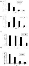
Mycoplasma hyopneumoniae is considered the major causative agent of porcine respiratory disease complex, occurs worldwide and causes major economic losses to the pig industry. To gain more insights into the pathogenesis of this organism, the high throughput cDNA microarray assays were employed to evaluate host responses of porcine alveolar macrophages to M. hyopneumoniae infection. A total of 1033 and 1235 differentially expressed genes were identified in porcine alveolar macrophages in responses to exposure to M. hyopneumoniae at 6 and 15 hours post infection, respectively. The differentially expressed genes were involved in many vital functional classes, including inflammatory response, immune response, apoptosis, cell adhesion, defense response, signal transduction, protein folding, protein ubiquitination and so on. The pathway analysis demonstrated that the most significant pathways were the chemokine signaling pathway, Toll-like receptor signaling pathway, RIG-I-like receptor signaling pathway, nucleotide-binding oligomerization domains (Nod)-like receptor signaling pathway and apoptosis signaling pathway. The reliability of the data obtained from the microarray was verified by performing quantitative real-time PCR. The expression kinetics of chemokines was further analyzed. The present study is the first to document the response of porcine alveolar macrophages to M. hyopneumoniae infection. The data further developed our understanding of the molecular pathogenesis of M. hyopneumoniae.
Conflict of interest statement
Figures


References
-
- Maes D, Verdonck M, Deluyker H, Kruif A (1996) Enzootic pneumonia in pigs. Vet Q 18: 104–9. - PubMed
-
- Thacker E (2006) Mycoplasmal diseases. The Iowa State University Press, Ames, IA, pp. 701–717.
-
- Blanchard B, Vena M, Cavalier A, Lannic J, Gouranton J, et al. (1992) Electron microscopic observation of the respiratory tract of SPF piglets inoculated with Mycoplasma hyopneumoniae . Vet Microbiol 30: 329–341. - PubMed
-
- Sarradell J, Andrada M, Ramairez AS, Fernandez A, Gaomez JC, et al. (2003) A morphologic and immunohistochemical study of the bronchus-associated lymphoid tissue of pigs naturally infected with Mycoplasma hyopneumoniae . Vet Pathol 40: 395–404. - PubMed
-
- Baskerville A (1972) Development of the early lesions in experimental enzootic pneumonia of pig: an ultrastructural and histological study. Res Vet Sci 13: 570–578. - PubMed
Publication types
MeSH terms
LinkOut - more resources
Full Text Sources
Other Literature Sources
Molecular Biology Databases

