. 2014 Dec;178(3):548-60.
doi: 10.1111/cei.12432.

Toll-like receptor 3 stimulation promotes Ro52/TRIM21 synthesis and nuclear redistribution in salivary gland epithelial cells, partially via type I interferon pathway

Affiliations

Toll-like receptor 3 stimulation promotes Ro52/TRIM21 synthesis and nuclear redistribution in salivary gland epithelial cells, partially via type I interferon pathway

N C Kyriakidis et al. Clin Exp Immunol. 2014 Dec.

Abstract

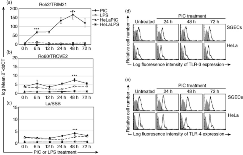
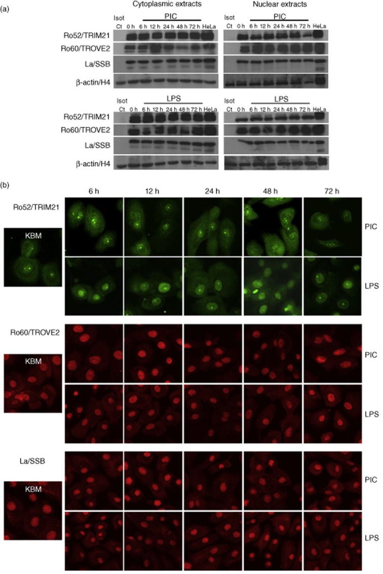
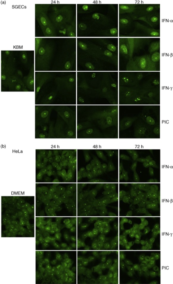
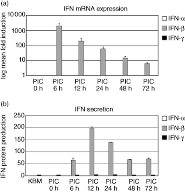
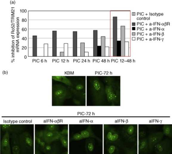
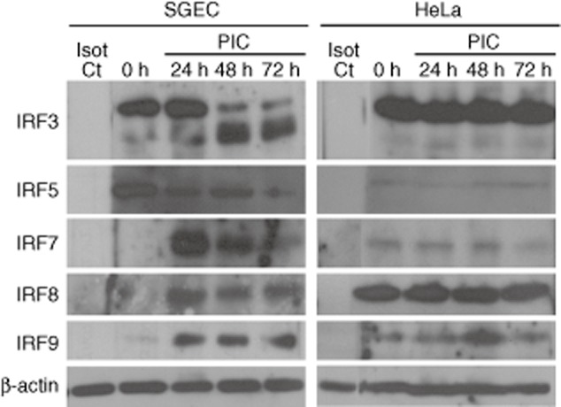
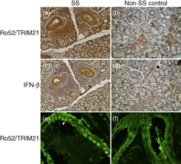
Up-regulated expression of Ro52/tripartite motif-containing protein 21 (TRIM21), Ro60/TROVE domain family, member 2 (TROVE2) and lupus LA protein/Sjögren's syndrome antigen B (La/SSB) autoantigens has been described in the salivary gland epithelial cells (SGEC) of patients with Sjögren's syndrome (SS). SGECs, the key regulators of autoimmune SS responses, express high levels of surface functional Toll-like receptor (TLR)-3, whereas Ro52/TRIM21 negatively regulates TLR-3-mediated inflammation. Herein, we investigated the effect of TLR-3-signalling on the expression of Ro52/TRIM21, as well as Ro60/TROVE2 and La/SSB autoantigens, by SGECs. The effect of TLR-3 or TLR-4 stimulation on autoantigen expression was evaluated by polyI:C or lipopolysaccharide (LPS) treatment, respectively, of SGEC lines (10 from SS patients, 12 from non-SS controls) or HeLa cells, followed by analysis of mRNA and protein expression. PolyI:C, but not LPS, resulted in a two-step induction of Ro52/TRIM21 mRNA expression by SGECs, a 12-fold increment at 6 h followed by a 2.5-fold increment at 24-48 h, whereas it induced a late two-fold up-regulation of Ro60/TROVE2 and La/SSB mRNAs at 48 h. Although protein expression levels were not affected significantly, the late up-regulation of Ro52/TRIM21 mRNA was accompanied by protein redistribution, from nucleolar-like pattern to multiple coarse dots spanning throughout the nucleus. These late phenomena were mediated significantly by interferon (IFN)-β production, as attested by cognate secretion and specific inhibition experiments and associated with IFN regulatory factor (IRF)3 degradation. TLR-3-signalling had similar effects on SGECs obtained from SS patients and controls, whereas it did not affect the expression of these autoantigens in HeLa cells. TLR-3 signalling regulates the expression of autoantigens by SGECs, implicating innate immunity pathways in their over-expression in inflamed tissues and possibly in their exposure to the immune system.

Keywords: Ro52/TRIM21 autoantigen; Sjögren's syndrome; TLR-3; salivary gland epithelial cells; type I interferons.


Figures

Fig. 1
Fig. 1
(a–c) Mean fold increase of mRNA expression following polyI:C (PIC) or lipopolysaccharide (LPS) treatment of salivary gland epithelial cells (SGECs) or HeLa cells. In SGECs, tripartite motif-containing protein 21 (TRIM21) mRNA (a) induction was evident from 6 h of polyI:C treatment (P < 0·0001), remained stable until 12–24 h and raised 2·5-fold at 48 h (compared to 12 h, P < 0·0001) (bold continuous line). LPS did not affect Ro52/TRIM21 mRNA expression (discontinuous line). Neither PolyI:C (thin continuous line) nor LPS (dotted line) affected Ro52/TRIM21 mRNA expression by HeLa cells. Treatment of SGECs with polyI:C was found to result in a slight, but statistically significant (P = 0·0002), induction of Ro60/TROVE domain family, member 2 (TROVE2) mRNA (b) at 48 h (compared to untreated, bold continuous line), whereas LPS had no effect (discontinuous line). Ro60/TROVE2 mRNA expression in HeLa cells was not affected by polyI:C (thin continuous line) or LPS (dotted line) treatment. Similarly, treatment of SGECs with polyI:C caused a slight, but statistically significant (P = 0·0014), lupus LA protein/Sjögren syndrome antigen B (La/SSB) mRNA (c) induction at 48 h (compared to untreated, bold continuous line), whereas LPS had no effect (discontinuous line). In HeLa cells, neither polyI:C (thin continuous line) nor LPS (dotted line) affected La/SSB mRNA expression. Standard errors are representative of all 22 SGEC lines and three different experiments on HeLa cells, whereas statistical significance is indicated by asterisks (***P < 0·001). Please note the different scale of the y-axes of the graphs. (d,e) Flow cytometric analysis of the surface Toll-like receptor (TLR)-3 (d) and TLR-4 (e) expression by untreated or polyI:C-treated Hela cells revealed significant constitutive expression of these receptors in both SGEC and HeLa cells, which is not affected significantly by TLR-3 stimulation.
Fig. 2
Fig. 2
PolyI:C treatment does not affect the levels of tripartite motif-containing protein 21 (TRIM21), Ro60/TROVE domain family, member 2 (TROVE2) or lupus LA protein/Sjögren syndrome antigen B (La/SSB) protein expression, but induces a late redistribution of Ro52/TRIM21 in the nucleus of salivary gland epithelial cells (SGECs). (a) Immunoblotting analysis did not reveal any notable changes in protein expression levels of Ro52/TRIM21, Ro60/TROVE2 and La/SSB in either cytoplasmic (left side) or nuclear (right side) extracts of SGECs upon polyI:C (PIC; upper panel) or lipopolysaccharide (LPS) (lower panel) treatment. β-actin and histone-H4 were used as loading controls for cytoplasmic and nuclear extracts, respectively. A representative example of seven SGEC lines is shown. (b) Confocal microscopy analysis revealed that polyI:C treatment induced a nuclear redistribution of Ro52/TRIM21 protein in SGECs. In untreated cells, Ro52/TRIM21 is localized at the cytoplasm and one or two nuclear dots, resembling nucleolar staining. This pattern of nuclear expression remained stable until 24 h, whereas at 48 and 72 h it was redistributed to multiple coarse dots spanning the nucleus. LPS treatment had no effect on Ro52/TRIM21, Ro60/TROVE2 and La/SSB protein expression or distribution. A representative example of 13 SGEC lines (eight from SS and five from non-SS controls) is shown.
Fig. 3
Fig. 3
Treatment with interferon (IFN)-α, IFN-β or IFN-γ induces the nuclear redistribution of tripartite motif-containing protein 21 (TRIM21) protein in salivary gland epithelial cells (SGECs), but not in HeLa cells. (a) IFN treatment of SGECs led to the nuclear redistribution of Ro52/TRIM21 protein from nucleolar-like pattern to multiple coarse dots spanning throughout the nucleus from 24 h. Representative example of three SGEC lines is shown. (b) Treatment with IFN-α, IFN-β or IFN-γ did not significantly alter the expression of Ro52/TRIM21 protein in HeLa cells. Figures are representative of three different experiments.
Fig. 4
Fig. 4
Production of interferons (IFN) by salivary gland epithelial cells (SGECs). (a) Histogram indicating the mRNA production of IFN-α, IFN-β and IFN-γ by resting or polyI:C (PIC)-treated SGECs. Treatment with polyI:C readily induced the expression of IFN-β, but not IFN-α or IFN-γ, mRNA in SGECs at 6 h, and this expression declined thereafter. (b) Histogram indicating the secretion of IFNs by resting or polyI:C-treated SGECs. Treatment with polyI:C induced IFN-β secretion from 6 h, with a peak at 12 h. Minute stable amounts of IFN-γ were detected in resting (0 h), polyI:C-treated SGECs and unused cultured medium (KBM), whereas IFN-α was not detected. Standard errors correspond to results from three SGEC lines.
Fig. 5
Fig. 5
Blockade of interferons (IFNs) by specific neutralizing antibodies revealed that the polyI:C-induced up-regulation of tripartite motif-containing protein 21 (TRIM21) mRNA and protein redistribution is mediated significantly by IFN-β. (a) Histogram showing the inhibition of Ro52/TRIM21 mRNA expression in resting or polyI:C (PIC)-treated salivary gland epithelial cells (SGECs) by specific neutralizing antibodies against IFN-α, IFN-β, IFN-γ, the common IFN-αβ-receptor (IFN-αβR) or isotype control. Antibodies against IFN-β or the common IFN-αβ-receptor significantly inhibit the polyI:C-driven mRNA increment, an effect that is more evident at the late increment (12–48 h; red box). (b) Blocking of polyI:C-induced redistribution of Ro52/TRIM21 protein by neutralizing antibodies against IFN-α, IFN-β, IFN-γ or IFN-αβ-receptor. Antibodies against IFN-β and IFN-αβ-receptor, but not against IFN-α or IFN-γ, block the polyI:C-driven nuclear redistribution of Ro52/TRIM21 in SGECs. Figures of blocking experiments at SGECs treated with polyI:C for 72 h are shown. These are representative of three distinct experiments.
Fig. 6
Fig. 6
Effect of polyI:C treatment in the expression of interferon regulatory factors (IRFs) by salivary gland epithelial cells (SGECs) and HeLa cells. Late polyI:C treatment resulted in the degradation of IRF3 in SGECs, but not in HeLa cells. Furthermore, stimulation of SGECs with polyI:C led to reduction of IRF5 expression, whereas it induced IRF7, IRF8 and IRF9 expression from 24 h. In line with mRNA and protein findings, IRF expression in HeLa cells was not affected by polyI:C treatment. β-actin was used as loading control. A representative example of four SGEC lines (two from SS and two from non-SS controls) is shown.
Fig. 7
Fig. 7
Representative imunohistochemical (a–d) and confocal microscopy (e,f) detection of tripartite motif-containing protein 21 (TRIM21) (a,b,e,f) and interferon (IFN)-β (c,d) expression at the minor salivary glands (MSG). Ro52/TRIM21 was found to be expressed strongly in the ductal and acinar epithelial cells of Sjögren's syndrome (SS) patients (a) and controls (b), as well as in the infiltrating mononuclear cells of SS patients (a). The IFN-β staining was significantly more intense at the ductal epithelial cells of SS patients (c) compared to non-SS controls (d). Confocal microscopy revealed that the majority of the ductal epithelial cells at the MSGs of SS patients present a coarse nuclear Ro52/TRIM21 staining pattern (arrow) (e). In contrast, approximately half the ductal epithelial cells at the MSGs of non-SS controls display nuclear Ro52/TRIM21 expression, which is more homogeneous than that of SS patients (f).

References

    1. Routsias JG, Vlachoyiannopoulos PG, Tzioufas AG. Autoantibodies to intracellular autoantigens and their B-cell epitopes: molecular probes to study the autoimmune response. Crit Rev Clin Lab Sci. 2006;43:203–248. - PubMed
    1. Routsias JG, Kyriakidis N, Latreille M, Tzioufas AG. RNA recognition motif (RRM) of La/SSB: the bridge for interparticle spreading of autoimmune response to U1-RNP. Mol Med. 2010;16:19–26. - PMC - PubMed
    1. Yiannaki E, Vlachoyiannopoulos PG, Manoussakis MN, et al. Study of antibody and T cell responses in rabbits immunized with synthetic human B cell epitope analogues of La (SSB) autoantigen. Clin Exp Immunol. 2000;121:551–556. - PMC - PubMed
    1. Tengner P, Halse AK, Haga HJ, Jonsson R, Wahren-Herlenius M. Detection of anti-Ro/SSA and anti-La/SSB autoantibody-producing cells in salivary glands from patients with Sjogren's syndrome. Arthritis Rheum. 1998;41:2238–2248. - PubMed
    1. Tzioufas AG, Hantoumi I, Polihronis M, Xanthou G, Moutsopoulos HM. Autoantibodies to La/SSB in patients with primary Sjogren's syndrome (pSS) are associated with upregulation of La/SSB mRNA in minor salivary gland biopsies (MSGs) J Autoimmun. 1999;13:429–434. - PubMed

Publication types