Review
. 2014 Apr 15;16(2):208.
doi: 10.1186/ar4542.

Molecular imaging of rheumatoid arthritis: emerging markers, tools, and techniques

Review

Molecular imaging of rheumatoid arthritis: emerging markers, tools, and techniques

Stéphanie Put et al. Arthritis Res Ther. .

Abstract

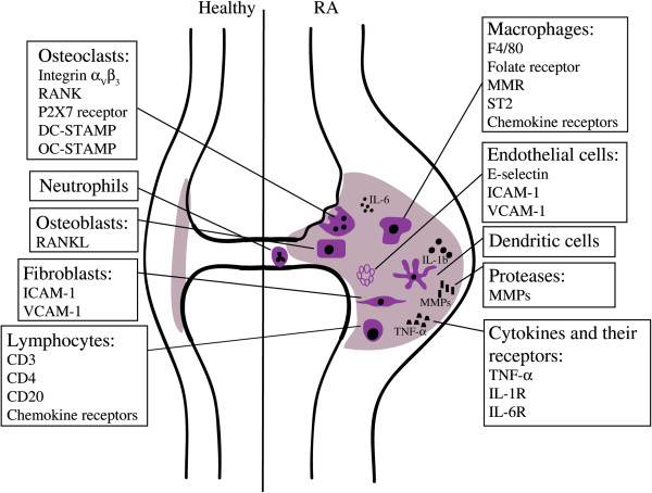
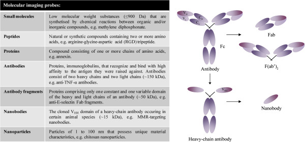
Early diagnosis and effective monitoring of rheumatoid arthritis (RA) are important for a positive outcome. Instant treatment often results in faster reduction of inflammation and, as a consequence, less structural damage. Anatomical imaging techniques have been in use for a long time, facilitating diagnosis and monitoring of RA. However, mere imaging of anatomical structures provides little information on the processes preceding changes in synovial tissue, cartilage, and bone. Molecular imaging might facilitate more effective diagnosis and monitoring in addition to providing new information on the disease pathogenesis. A limiting factor in the development of new molecular imaging techniques is the availability of suitable probes. Here, we review which cells and molecules can be targeted in the RA joint and discuss the advances that have been made in imaging of arthritis with a focus on such molecular targets as folate receptor, F4/80, macrophage mannose receptor, E-selectin, intercellular adhesion molecule-1, phosphatidylserine, and matrix metalloproteinases. In addition, we discuss a new tool that is being introduced in the field, namely the use of nanobodies as tracers. Finally, we describe additional molecules displaying specific features in joint inflammation and propose these as potential new molecular imaging targets, more specifically receptor activator of nuclear factor κB and its ligand, chemokine receptors, vascular cell adhesion molecule-1, αVβ₃ integrin, P2X7 receptor, suppression of tumorigenicity 2, dendritic cell-specific transmembrane protein, and osteoclast-stimulatory transmembrane protein.


Figures

Figure 1
Figure 1
Schematic overview of specific cells and molecules that can be targeted in the rheumatic joint. The rheumatoid synovium is characterized by the influx of inflammatory cells and release of cytokines. Surface molecules that are expressed on these cells can be used as markers to target and visualize the different cell types in the inflamed joint. DC-STAMP, dendritic cell-specific transmembrane protein; ICAM-1, intercellular adhesion molecule-1; IL, interleukin; MMP, matrix metalloproteinase; MMR, macrophage mannose receptor; OC-STAMP, osteoclast-stimulatory transmembrane protein; RA, rheumatoid arthritis; RANK, receptor activator of nuclear factor-kappa-B; RANKL, receptor activator of nuclear factor-kappa-B ligand; ST2, suppression of tumorigenicity 2; TNF-α, tumor necrosis factor-alpha; VCAM-1, vascular cell adhesion molecule-1.
Figure 2
Figure 2
Probes can be composed of small molecules, peptides, proteins, antibodies, antibody fragments, nanobodies, and nanoparticles. A schematic overview of a conventional antibody, a heavy-chain antibody, Fab fragments, and a nanobody is given. CH, heavy chain constant domain; CL, light chain constant domain; Fab, antigen-binding domain; Fc, constant domain; MMR, macrophage mannose receptor; TNF-α, tumor necrosis factor-alpha; VH, heavy chain variable domain; VHH, heavy chain only antibody VL, light chain variable domain.
Figure 3
Figure 3
Bone scintigraphy in a patient with active rheumatoid arthritis. Imaging was performed at 3 hours after injection of 740 MBq 99mtechnetium-methylene diphosphonate. The image shows increased tracer uptake in the wrists and joints of the fingers. The highest intensity is found in metacarpophalangeal joints.
Figure 4
Figure 4
In vivo imaging with macrophage mannose receptor (MMR)-specific nanobodies visualizes MMR expression in joints of mice with collagen-induced arthritis. Single-photon emission computed tomography and micro-computed tomography imaging was performed at 3 hours after injection of 99mtechnetium-labeled MMR-targeting nanobodies in mice without clinical symptoms of arthritis (A) (asymptomatic mice) or mice with arthritic joints (B) (symptomatic mice). Nanobodies against the β-lactamase BCII enzyme of Bacillus cereus (BCII10) were used as a non-targeting control. MMR staining is apparent in knees, ankles, and metatarsal joints of symptomatic mice (arrows) in addition to the signal in lymph nodes, liver, and spleen that is also detectable in asymptomatic mice. This image was originally published in the Journal of Nuclear Medicine[72]. © by the Society of Nuclear Medicine and Molecular Imaging, Inc.

References

    1. Mountz JM, Alavi A, Mountz JD. Emerging optical and nuclear medicine imaging methods in rheumatoid arthritis. Nat Rev Rheumatol. 2012;8:719–728. doi: 10.1038/nrrheum.2012.148. - DOI - PubMed
    1. Conti F, Malviya G, Ceccarelli F, Priori R, Iagnocco A, Valesini G, Signore A. Role of scintigraphy with mTc-infliximab in predicting the response of intraarticular infliximab treatment in patients with refractory monoarthritis. Eur J Nucl Med Mol Imaging. 2012;39:1339–1347. doi: 10.1007/s00259-012-2133-9. - DOI - PubMed
    1. Firestein GS. Evolving concepts of rheumatoid arthritis. Nature. 2003;423:356–361. doi: 10.1038/nature01661. - DOI - PubMed
    1. McInnes IB, Schett G. The pathogenesis of rheumatoid arthritis. N Engl J Med. 2011;365:2205–2219. doi: 10.1056/NEJMra1004965. - DOI - PubMed
    1. Cascao R, Rosario HS, Souto-Carneiro MM, Fonseca JE. Neutrophils in rheumatoid arthritis: more than simple final effectors. Autoimmun Rev. 2010;9:531–535. doi: 10.1016/j.autrev.2009.12.013. - DOI - PubMed