Review
. 2014 Aug;20(4 Neuro-ophthalmology):966-80.
doi: 10.1212/01.CON.0000453309.44766.b4.

Diplopia due to ocular motor cranial neuropathies

Review

Diplopia due to ocular motor cranial neuropathies

Wayne T Cornblath. Continuum (Minneap Minn). 2014 Aug.

Abstract

Purpose of review: Determining which cranial nerve(s) is (are) involved is a critical step in appropriately evaluating a patient with diplopia.

Recent findings: New studies have looked at the various etiologies of cranial nerve palsies in the modern imaging era. The importance of the C-reactive protein test in evaluating the possibility of giant cell arteritis has recently been emphasized.

Summary: Dysfunction of the oculomotor (third), trochlear (fourth), or abducens (sixth) cranial nerve will produce ocular misalignment and resultant binocular diplopia or binocular blur. A misalignment in the vertical plane of as small as 200 μm is enough to produce diplopia. Diagnosing diplopia from a cranial nerve abnormality requires an understanding of structure (the anatomy of the cranial nerves from nucleus to muscle), function (the movements controlled by the cranial nerves), possible etiologies, and exceptions to the rules.


Figures
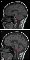
Figure 9-1.
Figure 9-1.
Panel A shows normal high signal in the clivus (arrow) on a T1-weighted MRI sagittal MRI of a healthy individual. In panel B, the normal high signal, from fat, in the clivus marrow space is absent in the T1-weighted sagittal MRI of the patient in Case 9-1 (arrow), indicating a metastatic process replacing the fat and compressing the sixth nerve in the Dorello canal.
Figure 9-2.
Figure 9-2.
Examination of the patient in Case 9-2. Normal right gaze (A), left ptosis and miosis (B), and abduction deficit on left gaze (C).
Figure 9-3.
Figure 9-3.
Testing with cocaine eye drops showed increase in anisocoria with minimal dilation on the left, indicating a left Horner syndrome.
Figure 9-4.
Figure 9-4.
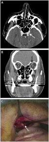
Imaging and biopsy of the patient in Case 9-2. A, Fullness of left cavernous sinus on axial CT (arrow). B, Enlarged superior orbital nerve on coronal CT (arrow). C, Lid crease incision and biopsy of enlarged superior orbital nerve seen on CT (arrow).
Figure 9-5.
Figure 9-5.
Examination of the patient in Case 9-3. A, Upgaze with slight limitation of elevation in the right eye. B, Right gaze. C, Primary gaze with right ptosis and pseudoretraction on left. D, Left gaze with decreased adduction of right eye. E, Downgaze with slight limitation of depression in the right eye.
Figure 9-6.
Figure 9-6.
Reconstruction of CT angiography showing right posterior communicating artery aneurysm (arrow).
Figure 9-7.
Figure 9-7.
Examination of the patient in Case 9-4. A, Upgaze showing lack of elevation of the left eye and adduction of the left eye. B, Right gaze showing slight limitation of adduction of the left eye. C, Primary gaze showing left ptosis. D, Left gaze showing dilation of the left pupil. E, Downgaze showing left eyelid retraction.
Figure 9-8.
Figure 9-8.
Axial contrast-enhanced T1-weighted MRI showing a large enhancing cavernous sinus meningioma on the left.
Figure 9-9.
Figure 9-9.
Examination of the patient in Case 9-5. A, Right gaze showing narrowed palpebral fissure in the left eye. B, Primary gaze showing normal alignment. C, Left gaze showing very limited abduction of the left eye.

References

    1. Hoya K,, Kirino T. Traumatic trochlear nerve palsy following minor occipital impact—four case reports. Neurol Med Chir (Tokyo) 2000; 40 (7): 358–360. - PubMed
    1. Trobe JD. Third nerve palsy and the pupil. Footnotes to the rule. Arch Ophthalmol 1988; 106 (5): 601–602. - PubMed
    1. Kissel JT,, Burde RM,, Klingele TG,, Zeiger HE. Pupil-sparing oculomotor palsies with internal carotid-posterior communicating artery aneurysms. Ann Neurol 1983; 13 (2): 149–154. - PubMed
    1. Dhume KU,, Paul KE. Incidence of pupillary involvement, course of anisocoria and ophthalmoplegia in diabetic oculomotor nerve palsy. Indian J Ophthalmol 2013; 61 (1): 13–17. - PMC - PubMed
    1. Schatz NJ,, Savino PJ,, Corbett JJ. Primary aberrant aculomotor regeneration. A sign of intracavernous meningioma. Arch Neurol 1977; 34 (1): 29–32. - PubMed