

Early intervertebral disc degeneration changes in asymptomatic weightlifters assessed by t1ρ-magnetic resonance imaging
- PMID: 25099319
- PMCID: PMC5585866
- DOI: 10.1097/BRS.0000000000000554
Early intervertebral disc degeneration changes in asymptomatic weightlifters assessed by t1ρ-magnetic resonance imaging
Abstract
Study design: Case-control study.
Objective: To evaluate early intervertebral disc degeneration quantified by T1ρ- and T2-weighted magnetic resonance imaging (MRI) in asymptomatic weightlifters compared with a healthy control group matched for sex and age.
Summary of background data: Athletes consistently recruit or transfer high levels of repetitive forces through the spine, and MRI has documented a higher rate of intervertebral disc degeneration in athletes compared with matched controls. This study aims to analyze the potential role of T1ρ-MRI in the assessment of early degenerative changes occurring in intervertebral discs of young asymptomatic weightlifters compared with healthy controls.
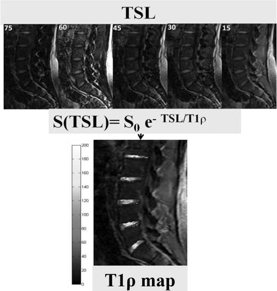
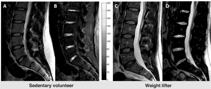
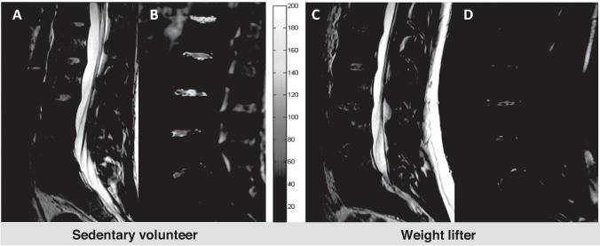
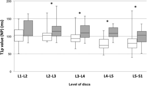
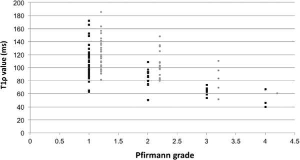
Methods: Twenty-six asymptomatic young male weightlifters versus a sedentary control group matched for age and sex, both having no lower back pain nor any spinal symptoms, underwent MRI (1.5 T). Degenerative grade was assessed using T2-weighted images, according to the Pfirrmann scale. T1ρ mapping and values in the nucleus pulposus (n=130) were obtained. Differences in T1ρ value between among the groups and linear regression analyses with degenerative grade were determined.
Results: Pfirrmann degenerative grade did not show significant differences among groups. Instead, T1ρ values were significantly lower in the lumbar spine of weightlifters compared with controls (P<0.05). T1ρ values decreased linearly with degenerative grade.
Conclusion: T1ρ values were significantly lower in athletes compared with a sedentary matched control group showing differences in intervertebral disc degeneration onset among individuals with lifestyle and environmental factors leading to back pain. T1ρ can be potentially used as a valid clinical tool to identify early changes in intervertebral disc on the verge of new emerging intervertebral discs regenerative strategies and treatments.
Level of evidence: 4.
Figures
Similar articles
-
T1ρ magnetic resonance imaging quantification of early lumbar intervertebral disc degeneration in healthy young adults.Spine (Phila Pa 1976). 2012 Jun 15;37(14):1224-30. doi: 10.1097/BRS.0b013e31824b2450. Spine (Phila Pa 1976). 2012. PMID: 22281486 Free PMC article.
-
T1ρ magnetic resonance imaging and discography pressure as novel biomarkers for disc degeneration and low back pain.Spine (Phila Pa 1976). 2011 Dec 1;36(25):2190-6. doi: 10.1097/BRS.0b013e31820287bf. Spine (Phila Pa 1976). 2011. PMID: 21358489 Free PMC article.
-
Glycosaminoglycan Chemical Exchange Saturation Transfer of Lumbar Intervertebral Discs in Healthy Volunteers.Spine (Phila Pa 1976). 2016 Jan;41(2):146-52. doi: 10.1097/BRS.0000000000001144. Spine (Phila Pa 1976). 2016. PMID: 26583472
-
Imaging Evaluation of Intervertebral Disc Degeneration and Painful Discs-Advances and Challenges in Quantitative MRI.Diagnostics (Basel). 2022 Mar 14;12(3):707. doi: 10.3390/diagnostics12030707. Diagnostics (Basel). 2022. PMID: 35328260 Free PMC article. Review.
-
T1ρ magnetic resonance: basic physics principles and applications in knee and intervertebral disc imaging.Quant Imaging Med Surg. 2015 Dec;5(6):858-85. doi: 10.3978/j.issn.2223-4292.2015.12.06. Quant Imaging Med Surg. 2015. PMID: 26807369 Free PMC article. Review.
Cited by
-
The association between occupational loading and spine degeneration on imaging - a systematic review and meta-analysis.BMC Musculoskelet Disord. 2019 Oct 27;20(1):489. doi: 10.1186/s12891-019-2835-2. BMC Musculoskelet Disord. 2019. PMID: 31656182 Free PMC article.
-
Comprehensive assessment of in vivo lumbar spine intervertebral discs using a 3D adiabatic T1ρ prepared ultrashort echo time (UTE-Adiab-T1ρ) pulse sequence.Quant Imaging Med Surg. 2022 Jan;12(1):269-280. doi: 10.21037/qims-21-308. Quant Imaging Med Surg. 2022. PMID: 34993077 Free PMC article.
-
Robotic Spine Surgery and Augmented Reality Systems: A State of the Art.Neurospine. 2020 Mar;17(1):88-100. doi: 10.14245/ns.2040060.030. Epub 2020 Mar 31. Neurospine. 2020. PMID: 32252158 Free PMC article.
-
Biomechanical Study of Symmetric Bending and Lifting Behavior in Weightlifter with Lumbar L4-L5 Disc Herniation and Physiological Straightening Using Finite Element Simulation.Bioengineering (Basel). 2024 Aug 12;11(8):825. doi: 10.3390/bioengineering11080825. Bioengineering (Basel). 2024. PMID: 39199783 Free PMC article.
-
Prevalence of degenerative vertebral disc changes in elite female Crossfit athletes - a cross-sectional study.BMC Musculoskelet Disord. 2023 Dec 11;24(1):963. doi: 10.1186/s12891-023-07071-9. BMC Musculoskelet Disord. 2023. PMID: 38082262 Free PMC article.
References
-
- d’Hemecourt PA, Gerbino PG, II, Micheli LJ. Back injuries in the young athlete. Clin Sports Med. 2000;19:663–79. - PubMed
-
- Graw BP, Wiesel SW. Low back pain in the aging athlete. Sports Med Arthrosc. 2008;16:39–46. - PubMed
-
- Taimela S, Kujala UM, Salminen JJ, et al. The prevalence of low back pain among children and adolescents. A nationwide, cohort-based questionnaire survey in Finland. Spine (Phila Pa 1976) 1997;22:1132–6. - PubMed
-
- Chan SC, Ferguson SJ, Wuertz K, et al. Biological response of the intervertebral disc to repetitive short-term cyclic torsion. Spine (Phila Pa 1976) 2011;36:2021–30. - PubMed
MeSH terms
Grants and funding
LinkOut - more resources
Full Text Sources
Other Literature Sources
Medical
Research Materials
