. 2014 Dec;5(6):715-22.
doi: 10.1007/s13244-014-0344-2. Epub 2014 Aug 7.

Breast imaging findings in haematological malignancies

Affiliations

Breast imaging findings in haematological malignancies

K N Glazebrook et al. Insights Imaging. 2014 Dec.

Abstract

Objectives: The objectives of this article are to review and illustrate the imaging appearances of haematological malignancies in the breast.

Methods: With Institutional Review Board approval, a search of the surgical pathology records from 1st January 2000 to 1st July 2012 was performed for haematological malignancies.

Results: Forty-eight cases of haematological malignancies (42 women and 6 men) were identified with imaging available for review: 39 cases of breast lymphoma, 6 cases of chronic lymphocytic leukaemia, 2 cases of acute leukaemia and 1 case of known multiple myeloma.

Conclusions: Breast manifestations of haematological malignancies are rare. They can have a variable appearance at imaging and can mimic primary breast carcinoma. In the setting of suspicious breast imaging findings, pathological diagnosis of haematological malignancy is concordant. Correlation with a clinical history of prior haematological malignancy can be helpful in suggesting the diagnosis and help prevent unnecessary surgical treatment.

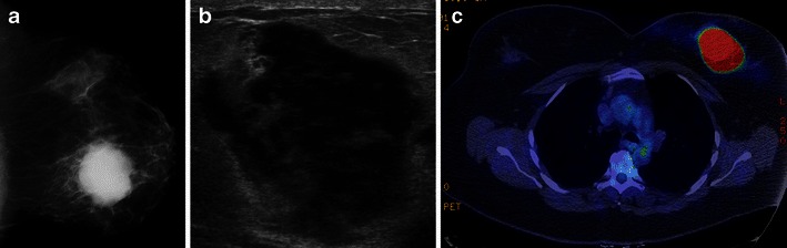
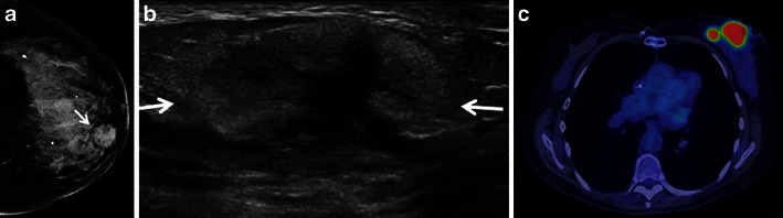
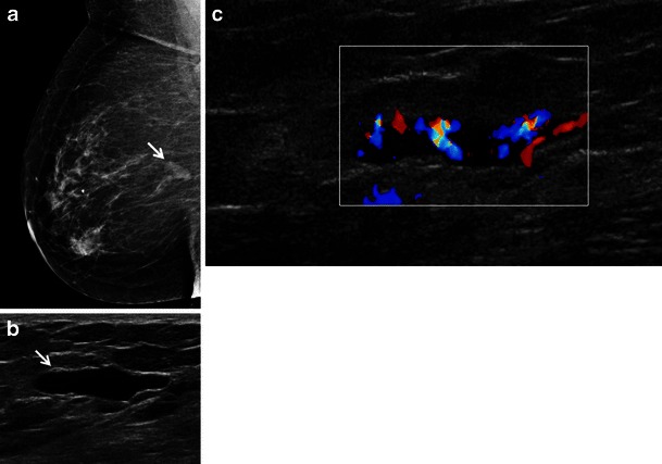
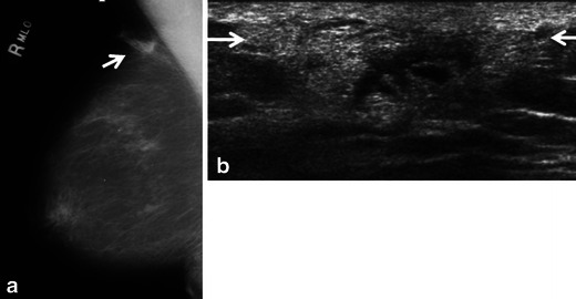
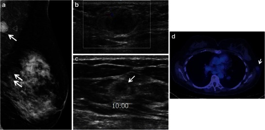
Teaching points: • Breast haematological malignancies are rare but the imaging appearances can mimic breast carcinoma. • Breast lymphoma, most often B-cell non-Hodgkin lymphoma, may be primary or due to secondary disease. • At ultrasound, haematological malignancies may present as a heterogeneous or predominantly echogenic mass. • Haematological malignancies show intense activity on PET/CT except myeloma which has low FDG uptake.


Figures

Fig. 1
Fig. 1
A 45-year-old woman with primary B-cell lymphoma presents with a palpable mass in the left breast. a Left cranio-caudal mammogram demonstrates a high-density irregular mass with indistinct margins in the medial left breast. b Ultrasound of the palpable mass demonstrates an irregular hypoechoic mass with thin echogenic rim. c Axial PET/CT shows homogeneous intense hypermetabolic activity within the mass. No uptake is seen within normal appearing nodes in the left axilla (not shown)
Fig. 2
Fig. 2
A 74-year-old woman with primary large B cell breast lymphoma presenting with a palpable mass in the left breast. a Cranio-caudal left breast mammogram shows an isodense circumscribed mass (arrow) in the medial left breast. b Ultrasound of the palpable mass demonstrates an oval, circumscribed principally echogenic mass with an irregular shaped central area of hypoechogenicity (arrows). c PET/CT shows two areas of homogeneous hypermetabolic uptake in the medial left breast. No axillary lymph node uptake was seen (not shown)
Fig. 3
Fig. 3
A 67-year-old woman with a history of marginal zone B-cell lymphoma (mucosa associated lymphoid tissue [MALT] lymphoma) presents with screening mammogram detected new mass in the right breast. a Right medio-lateral oblique mammogram demonstrates a lobulated circumscribed isodense mass in the upper right breast posterior depth (arrow). b, c Ultrasound shows an anechoic pseudocystic mass without posterior enhancement (arrow) which shows marked increased vascularity on colour Doppler evaluation indicating this is a solid mass
Fig. 4
Fig. 4
A 72-year-old woman with marginal zone B-cell lymphoma with follicular colonisation presents with developing focal asymmetry within the right upper breast noted on screening mammogram. a. Ill-defined focal asymmetry noted on the right medio-lateral oblique screening mammogram (arrow). b Ultrasound shows an irregular principally hyperechoic mass (arrows) extending to involve the skin with central areas of hypoechogenicity
Fig. 5
Fig. 5
A 73-year-old man with a history of CLL and a new palpable mass in the left breast with associated nipple retraction. Biopsy showed chronic lymphocytic leukaemia. a Left breast medio-lateral oblique and cranio-caudal mammograms show an oval circumscribed subareolar mass with mild nipple retraction (arrows). b, c Ultrasound demonstrates an irregularly shaped, hypoechoic mass with angular margins and a thick echogenic rim which shows increased vascularity on colour Doppler evaluation (arrows). d Strain elastography (red equating to hard) demonstrates a hard mass which is significantly larger on the elastogram. The ratio of size was 1.99 between the B-mode and strain image.
Fig. 6
Fig. 6
A 76-year-old man with known CLL presents with new palpable mass in the medial right breast which could not be included on a mammogram. Biopsy showed CLL. a Fused PET/CT image shows a hypermetabolic mass corresponding to the palpable mass in the medial right breast. b Fused PET/CT image demonstrates hypermetabolic axillary adenopathy (arrows). c Ultrasound shows a cluster of multiple hypoechoic masses with microlobulated margins without increased through transmission (arrows)
Fig. 7
Fig. 7
An 83-year-old woman with known acute T-cell prolymphocytic leukaemia presents with new palpable masses in the left breast. a Left medio-lateral oblique and cranio-caudal diagnostic mammogram demonstrates two areas of architectural distortion corresponding to the two palpable masses (arrows). b Ultrasound shows ill-defined mixed echogenic and hypoechoic mass (arrows). The other palpable mass had a similar sonographic appearance (not shown)
Fig. 8
Fig. 8
A 57-year-old woman with known multiple myeloma of the sacrum and multifocal involvement of the left breast and low axilla. a Full-field digital medio-lateral oblique view of the left breast demonstrates a round, circumscribed 1.9-cm mass (arrow) in the upper outer left breast/low axilla corresponding to a palpable abnormality. Two additional circumscribed masses were noted in the superior left breast posterior depth (arrows). b Ultrasound of the palpable low axillary mass imaging demonstrates a 2.0-cm heterogeneous mass with circumscribed margins with only mild increase in vascularity on colour Doppler. c Ultrasound of the mass at the 10 o’clock position of the left breast shows a microlobulated principally echogenic mass with central area of hypoechogenicity (arrow). The mass at the 9 o’clock position of the left breast had a similar sonographic appearance (not shown). Ultrasound guide core biopsy of all three lesions showed multiple myeloma. d Minimal FDG uptake is noted within the left upper outer/low axillary mass (arrow) on PET/CT

References

    1. American Cancer Society . Cancer facts & figures 2013. Atlanta: American Cancer Society; 2013.
    1. Shim E, Song SE, Seo BK, Kim Y-S, Son GS. Lymphoma affecting the breast: a pictorial review of multimodal imaging findings. J Breast Cancer. 2013;16(3):254–265. doi: 10.4048/jbc.2013.16.3.254. - DOI - PMC - PubMed
    1. Liberman L, Giess CS, Dershaw DD, Louie DC, Deutch BM. Non-Hodgkin lymphoma of the breast: imaging characteristics and correlation with histopathologic findings. Radiology. 1994;192:157–160. doi: 10.1148/radiology.192.1.8208929. - DOI - PubMed
    1. Wiseman C, Liao KT. Primary lymphoma of the breast. Cancer. 1972;29:1705–1712. doi: 10.1002/1097-0142(197206)29:6<1705::AID-CNCR2820290640>3.0.CO;2-I. - DOI - PubMed
    1. Paes FM, Kalkanis DG, Sideras PA, Serafini AN. FDG PET/CT of extranodal involvement in non-Hodgkin lymphoma and Hodgkin disease. Radiographics. 2010;30:269–291. doi: 10.1148/rg.301095088. - DOI - PubMed

LinkOut - more resources