. 2014 Oct 1;307(7):C648-56.
doi: 10.1152/ajpcell.00210.2014. Epub 2014 Aug 6.

Transcriptional regulation of α1H T-type calcium channel under hypoxia

Affiliations

Transcriptional regulation of α1H T-type calcium channel under hypoxia

Hassan Sellak et al. Am J Physiol Cell Physiol. .

Abstract

The low-voltage-activated T-type Ca(2+) channels play an important role in mediating the cellular responses to altered oxygen tension. Among three T-type channel isoforms, α1G, α1H, and α1I, only α1H was found to be upregulated under hypoxia. However, mechanisms underlying such hypoxia-dependent isoform-specific gene regulation remain incompletely understood. We, therefore, studied the hypoxia-dependent transcriptional regulation of α1G and α1H gene promoters with the aim to identify the functional hypoxia-response elements (HREs). In rat pulmonary artery smooth muscle cells (PASMCs) and pheochromocytoma (PC12) cells after hypoxia (3% O2) exposure, we observed a prominent increase in α1H mRNA at 12 h along with a significant rise in α1H-mediated T-type current at 24 and 48 h. We then cloned two promoter fragments from the 5'-flanking regions of rat α1G and α1H gene, 2,000 and 3,076 bp, respectively, and inserted these fragments into a luciferase reporter vector. Transient transfection of PASMCs and PC12 cells with these recombinant constructs and subsequent luciferase assay revealed a significant increase in luciferase activity from the reporter containing the α1H, but not α1G, promoter fragment under hypoxia. Using serial deletion and point mutation analysis strategies, we identified a functional HRE at site -1,173cacgc-1,169 within the α1H promoter region. Furthermore, an electrophoretic mobility shift assay using this site as a DNA probe demonstrated an increased binding activity to nuclear protein extracts from the cells after hypoxia exposure. Taken together, these findings indicate that hypoxia-induced α1H upregulation involves binding of hypoxia-inducible factor to an HRE within the α1H promoter region.

Keywords: T-type calcium channel; gene expression; hypoxia; hypoxia-response element.


Figures

Fig. 1.
Fig. 1.
Selective upregulation of α1H under hypoxia. A: rat pulmonary artery smooth muscle cells (PASMCs), pulmonary microvascular endothelial cells (PMVECs), PC12 cells, and A7r5 cells were subjected to hypoxia (3% O2) or maintained under normoxia (21% O2) for 12 h. RT-PCR amplification was performed using total RNA isolated from each of the cells at the end of the exposure with the primer sets indicated in materials and methods. B: immunofluorescence labeling of α1G and α1H in PASMCs and PC12 cells. Representative projected experimental images of all parts of z-stacks taken from apical to basal cell aspects at 0.2-μm intervals. Immunofluorescence labeling was performed with an anti-α1G or anti-α1H antibody followed by an Alexa Fluor 488- or 594-conjugated secondary antibody (green or red) and nuclei visualized with DAPI (blue). Note the increase in Alexa Fluor 488 and 594 fluorescence in PC12 cells and PASMCs after 48-h exposure to hypoxia (3% O2) (denoted by arrows or arrowheads). Images acquired at 21°C on a Nikon A1 confocal laser microscope with a ×60, 1.20 numerical aperture water immersion objective (replicated 3 times).
Fig. 2.
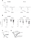
Fig. 2.
Functional expression of the α1H T-type Ca2+ channel is elevated under hypoxia. A: representative deactivating tail current traces from PASMCs and PC12 cells after 24-h or 48-h exposure to hypoxia (3% O2) (gray, ■) or under normoxia (black, □), elicited by repolarizing the cell to −120 mV following a short depolarizing pulse at +80 mV (3.25 ms) from a holding potential of −90 mV, as schematically illustrated. B: scatter plot of individual data points of maximally elicited T-type tail currents (Itail) from PASMCs and PC12 cells after 24-h or 48-h exposure to hypoxia or under normoxia (hypoxia: ■, normoxia: □; median with range: red lines). Note Itail median in PASMCs after hypoxia: −7.1 pA/pF vs. −2.3 pA/pF under normoxia at 24 h (n = 12 each, P < 0.05), −3.9 pA/pF vs. −2.2 pA/pF under normoxia at 48 h (n = 20 and 22, P < 0.001); in PC12 cells after hypoxia: 25.9 pA/pF vs. −16.3 pA/pF under normoxia at 24 h (n = 15 each, P < 0.05), −34.5 pA/pF vs. −18.4 pA/pF under normoxia at 48 h (n = 15 each, P < 0.05). C and D: comparison of the T-type currents in PASMCs after 24-h exposure to hypoxia (3% O2) to those in PASMCs maintained under normoxia (21% O2) for the same period of time (hypoxia: ■, normoxia: □). C: representative macroscopic current traces recorded a depolarization to −30 mV from a holding potential of −90 mV, as schematically illustrated. D: current-voltage (I–V) relationships of peak currents recorded by step depolarizations to varying test potentials (−70 to +50 mV with 10-mV increments) from a holding potential of −90 mV. Data represent the means ± SE, n = 12 each. Ca2+ (10 mmol/l) was used as a charge carrier. Nifedipine (10 μmol/l) was present in the extracellular solution. Temperature was 22–25°C.
Fig. 3.
Fig. 3.
The α1G promoter activity was unchanged under hypoxia. Serial deletion constructs of the 5′ flaking region of α1G were generated as described in materials and methods. PC12 cells were transiently cotransfected with different α1G serial deletion constructs (250 ng) and 50 ng of β-galactosidase using Fugene 6 HD (1 μl) as transfecting reagent. Cells were then exposed to hypoxia (3% O2) (H) or normoxia (21% O2) (N) for 48 h. Luciferase assays were performed as described in materials and methods, and values were normalized to β-galactosidase serving as internal control for transfection efficiency and expressed as relative light units (RLU). Representative of 3 independent experiments performed in triplicate. Left: potential hypoxia-response elements (HRE) on the 2,000-bp 5′ franking sequence of α1G: −1901gcgtg−1895 and −73gcgtg−69 on forward strand.
Fig. 4.
Fig. 4.
Hypoxia increases α1H promoter activity. PC12 cells were transiently cotransfected with different α1H serial deletion constructs (250 ng) and β-galactosidase (50 ng). Cells were then exposed to hypoxia (3% O2) for 48 h. Luciferase assays were performed as described in materials and methods. The luciferase values were normalized to β-galactosidase activity, which served as internal control for transfection efficiency. Representative of 3 independent experiments performed in triplicate. *P < 0.05, **P < 0.005. Left: potential HRE on the 3,076-bp 5′-flanking sequence of α1H: −3,025acgtg−3,021, −2,933acgtg−2,929, and −1,090gcgtg−1,086 on forward strand; −3,026cacgt−3,022, −2,934cacgt−2,930, −2,315cacgc−2,311, −1,182cacgt−1,178, −1,173cacgc−1,169, −1,132cacgc−1,128, −1,114cacgc−1,110, −1,079cacgc−1,075, and −1,079cacgc−1,075 on reverse strand. Arrow denotes the HRE site responsive to hypoxia.
Fig. 5.
Fig. 5.
HRE-5 within α1H promoter mediates hypoxia response. A: hypoxia increases hypoxia-inducible factor (HIF)-1α binding to HRE-5. PC12 cells and PASMCs were subjected to hypoxia (3% O2) for 6 h. Nuclear protein extracts were prepared as described in materials and methods and incubated with different radio-labeled probes consensus sequence (I), HRE-5 (II) within α1H promoter, mutated HRE-5 (III) for 30 min before resolving DNA-protein complex on gel. In the immune competition assay, the anti-HIF-1α antibody was incubated for 30 min with nuclear extracts prepared from PC12 subjected to hypoxia (3% O2 for 6 h) before addition of the probe. F0, free probe; N, normoxia (21% O2); H, hypoxia (3% O2). B: HRE-5 mutation abolishes hypoxia response. Site-directed mutagenesis was performed as described in materials and methods to generate constructs F1 and F5 harboring mutated HRE-5 named mF1 and mF5, respectively. PC12 cells were cotransfected with wild-type (F1, F5) or mutated HRE-5 (mF1, mF5) luciferase reporter constructs (250 ng) and β-galactosidase (50 ng). Cells were then exposed to hypoxia (3% O2 for 24 h). The luciferase values (RLU) were normalized to β-galactosidase activity serving as internal control. Representative of 3 independent experiments performed in triplicate. CACGC, wild-type HRE-5; CAAAC, mutated HRE; F0, empty vector. *P < 0.05, **P < 0.005.

References

    1. Akaike T, Jin MH, Yokoyama U, Izumi-Nakaseko H, Jiao Q, Iwasaki S, Iwamoto M, Nishimaki S, Sato M, Yokota S, Kamiya Y, Adachi-Akahane S, Ishikawa Y, Minamisawa S. T-type Ca2+ channels promote oxygenation-induced closure of the rat ductus arteriosus not only by vasoconstriction but also by neointima formation. J Biol Chem 284: 24025–24034, 2009 - PMC - PubMed
    1. Babal P, Manuel SM, Olson JW, Gillespie MN. Cellular disposition of transported polyamines in hypoxic rat lung and pulmonary arteries. Am J Physiol Lung Cell Mol Physiol 278: L610–L617, 2000 - PubMed
    1. Barrett PQ, Ertel EA, Smith MM, Nee JJ, Cohen CJ. Voltage-gated calcium currents have two opposing effects on the secretion of aldosterone. Am J Physiol Cell Physiol 268: C985–C992, 1995 - PubMed
    1. Becker AJ, Pitsch J, Sochivko D, Opitz T, Staniek M, Chen CC, Campbell KP, Schoch S, Yaari Y, Beck H. Transcriptional upregulation of CaV3.2 mediates epileptogenesis in the pilocarpine model of epilepsy. J Neurosci 28: 13341–13353, 2008 - PMC - PubMed
    1. Bertolesi GE, Jollimore CA, Shi C, Elbaum L, Denovan-Wright EM, Barnes S, Kelly ME. Regulation of α1G T-type calcium channel gene (cacna1g) expression during neuronal differentiation. Eur J Neurosci 17: 1802–1810, 2003 - PubMed

Publication types

LinkOut - more resources