

Comment
doi: 10.1016/j.bpj.2014.06.019.
Cross-talk, cross-bridges, and calcium activation of cardiac contraction
Affiliations
- PMID: 25099792
- PMCID: PMC4129475
- DOI: 10.1016/j.bpj.2014.06.019
Item in Clipboard
Comment
Cross-talk, cross-bridges, and calcium activation of cardiac contraction
Biophys J.
.
Erratum in
- Biophys J. 2014 Sep 16;107(6):1485
No abstract available
Figures
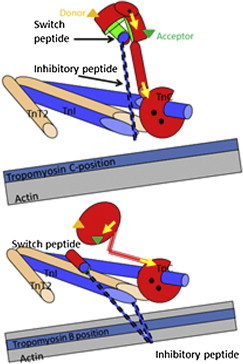
The thin-filament calcium switch. The Tn complex in the thin filament based on crystal structures of skeletal muscle TnC (red) with TnI (blue), and TnT2 (brown). This shows the role of the switch and inhibitory peptides of TnI in transmitting the calcium binding to TnC to the tropomyosin (pale blue strand) position on actin filament (gray strand). (Upper panel) Calcium-bound state. Calcium (black dots) binding to the N-terminal domain causes the regulatory domain to open, allowing the TnI signal peptide to bind in the cleft (the TnC and switch-peptide contact surface is shown in green). This inhibits the peptide from binding to its site on actin. Tm sits in its preferred C- or calcium-induced site on actin. (Lower panel) Calcium-free state with the inhibitory TnI peptide binding to actin and locating Tm in the Blocked position on actin. Note the closed regulatory domain of TnC. Note also that the diagram shows the role of the inhibitory peptide but not the rest of the C-terminus of TnI, which also contributes to the actin binding site. This is based on the crystal structure of skeletal muscle troponin core domain. The crystal structure of the cardiac troponin core domain does not show the central helix straightening as depicted here. This region may therefore be quite mobile. The locations of the fluorescent labels are for illustration only: (green and orange triangles) the FRET probes in the N-domain of TnC; (yellow arrows) the two bifunctional orientation probes in the two TnC domains. The figure is based on an original drawing by M. Vinogradova and R. Fletterick (3). To see this figure in color, go online.
Comment on
-
In situ time-resolved FRET reveals effects of sarcomere length on cardiac thin-filament activation.Biophys J. 2014 Aug 5;107(3):682-693. doi: 10.1016/j.bpj.2014.05.044. Biophys J. 2014. PMID: 25099807 Free PMC article.
References
-
- Lakatta E.G. Starling’s law of the heart is explained by an intimate interaction of muscle length and myofilament calcium activation. J. Am. Coll. Cardiol. 1987;10:1157–1164. - PubMed
-
- Geeves M.A. Thin filament regulation. In: Egelman E.H., editor. Vol. 4. Academic Press, Elsevier; Amsterdam, The Netherlands: 2012. pp. 251–267. (Comprehensive Biophysics).
-
- Gordon A.M., Homsher E., Regnier M. Regulation of contraction in striated muscle. Physiol. Rev. 2000;80:853–924. - PubMed
-
- Dong W.J., Robinson J.M., Cheung H.C. Kinetics of conformational transitions in cardiac troponin induced by Ca2+ dissociation determined by Förster resonance energy transfer. J. Biol. Chem. 2003;278:42394–42402. - PubMed
Publication types
MeSH terms
Substances
LinkOut - more resources
Full Text Sources
Other Literature Sources
