. 2014 Aug 5;107(3):599-612.
doi: 10.1016/j.bpj.2014.05.037.

Characterization of a novel water pocket inside the human Cx26 hemichannel structure

Affiliations

Characterization of a novel water pocket inside the human Cx26 hemichannel structure

Raul Araya-Secchi et al. Biophys J. .

Abstract

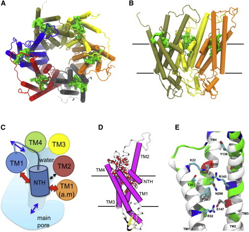
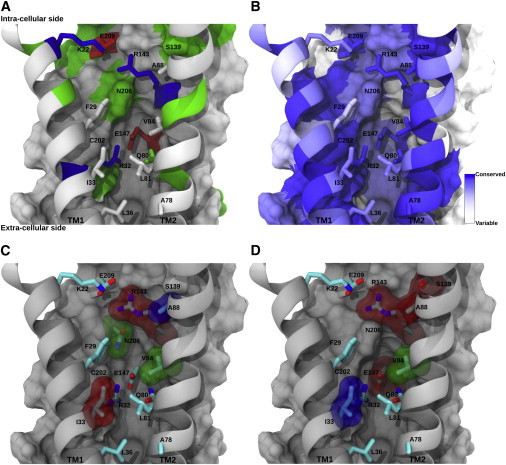
Connexins (Cxs) are a family of vertebrate proteins constituents of gap junction channels (GJCs) that connect the cytoplasm of adjacent cells by the end-to-end docking of two Cx hemichannels. The intercellular transfer through GJCs occurs by passive diffusion allowing the exchange of water, ions, and small molecules. Despite the broad interest to understand, at the molecular level, the functional state of Cx-based channels, there are still many unanswered questions regarding structure-function relationships, perm-selectivity, and gating mechanisms. In particular, the ordering, structure, and dynamics of water inside Cx GJCs and hemichannels remains largely unexplored. In this work, we describe the identification and characterization of a believed novel water pocket-termed the IC pocket-located in-between the four transmembrane helices of each human Cx26 (hCx26) monomer at the intracellular (IC) side. Using molecular dynamics (MD) simulations to characterize hCx26 internal water structure and dynamics, six IC pockets were identified per hemichannel. A detailed characterization of the dynamics and ordering of water including conformational variability of residues forming the IC pockets, together with multiple sequence alignments, allowed us to propose a functional role for this cavity. An in vitro assessment of tracer uptake suggests that the IC pocket residue Arg-143 plays an essential role on the modulation of the hCx26 hemichannel permeability.


Figures

Figure 1
Figure 1
Overview of the IC pocket. (A) View from the intracellular side of the hCx26 hemichannel. The IC pocket location can be depicted by the presence of a green solid volume in every monomer. (B) View from inside the pore of the hCx26 hemichannel showing only three monomers for clarity. Protein is rendered in cartoon according to the secondary structure and colored by monomer. Water molecules inside each IC pocket are shown as a green solid volume (A) and (B). In (B) and (C), black solid lines depict the boundaries of the lipid membrane according to OPM database (see Methods). (C) Cartoon representation of one hCx26 monomer from a side view. Protein has been colored by secondary structure (magenta = α-helix; yellow = β-sheet; white = loop) and water molecules filling the IC pocket are shown in vdW representation colored by element (red = oxygen; white = hydrogen). (D) Schematic overview of the IC pocket location. Depicted cartoon represents a view from the intracellular side of one hCx26 monomer composing a hemichannel. Each circle represents a TM-helix. The orange circle represents TM1 from the clockwise adjacent monomer (a.m.). The blue cylinder represents NTH and the red and black arrows depict relevant interactions between NTH and residues from the same or the adjacent monomer. Blue arrows indicate the water entry and exit points. (E) Selected snapshot from the MD simulation depicting relevant residues forming the IC pocket. Protein is rendered as cartoon representation and colored by residue type (red = acidic; blue = basic; green = polar; white = nonpolar). Side chains are shown in licorice representation and colored by element (red = oxygen; blue = nitrogen; cyan = carbon). Hydrogen atoms and residues 1 to 14 (NTH) were omitted for clarity. To see this figure in color, go online.
Figure 2
Figure 2
Conservation of residues in the IC pocket. (A) General overview of the IC pocket where constituent residues are rendered using licorice and color-coded by residue in equivalent orientation to Fig. 1E. Buried residues belonging to the IC pocket appear as colored surfaces. Transmembrane helices TM1 and TM2 are rendered using ribbons. (B) Residues belonging to the IC pocket, color-coded according to the ConSurf conservation score that appears at the bottom left. (C) IC pocket residues with associated mutations reported in the literature (see Table 2). The vdW representation is color-coded according to the functionality of the resulting mutant hCx26 hemichannel (red = diminished or null function; green = permeability and selectivity changes; blue = depending on the study may be classified red or green). (D) Mapping on the IC pocket of the amino acid residues reported by Skerret et al. (67). Residues surrounded by a transparent surface correspond to those that were mutated to cysteine showing relevant effects. Residues marked in red (R143, A88, S139, and E147) produced hemichannels with altered gating properties but normal conductance. Green-colored residues (V84) produced hemichannels with a diminished conductance. Blue-colored residues (I33) produced hemichannels with significant reduction in conductance. To see this figure in color, go online.
Figure 3
Figure 3
Water dynamics inside the IC pocket of each hCx26 monomer. (A) Water occupancy depicted as a box plot showing the number of water molecules inside the IC pocket of each Cx26 monomer. Box boundaries represent the standard deviation from the average position marked by the small square. The central line on each box shows the median value. Whiskers represent percentile 5 (top) and 95 (bottom) and the stars denote maximum (top) and minimum (bottom) values. Data were collected from four independent MD simulations of 20 ns each (see Methods). (B) Survival probabilities P(t) for water molecules inside the IC pocket of each hCx26 monomer. Data was taken averaging per monomer for each independent MD simulation of 20 ns. Each P(t) was obtained by averaging for each of the time windows available at the given interval (from 1 to 50 ps). Error bars represent the standard deviation from the block-average depicted in solid and using monomer-colored lines. To see this figure in color, go online.
Figure 4
Figure 4
Orientational time correlation of water molecules within the IC pocket. (A) Correlation time C2,û(t) for ûdipole. (B) Correlation time C2,û(t) for ûOH. In both panels correlation time values obtained for bulk water are shown as a gray-dashed line. Data shown using solid lines correspond to the average obtained from each data point between the four independent 20 ns MD production runs. Error bars represent the standard deviation for every average point. To see this figure in color, go online.
Figure 5
Figure 5
Arg-143 may adopt three main conformations on the IC pocket. (AC) Probability distribution of distances between Arg-143:Cζ and Glu-147:C; Arg-143:Cζ and Glu-209:C; and Arg-143:Cζ and center of mass of the phenyl ring of Phe-29, respectively, for each hCx26-monomer. (D) Panel shows representative conformations found for Arg-143 and Phe-29 from all six monomers along the MD production run. All monomers were aligned against one monomer from the crystal structure to observe, in one IC pocket, all the conformations adopted by Arg-143 and Phe-29. Other residues from the IC pocket are represented as a solid surface and colored by residue type (blue = positive; red = negative; green = polar; white = hydrophobic). Residues Arg-143 and Phe-29 are shown in licorice representation colored by monomer (black = M1; red = M2; green = M3; blue = M4; cyan = M5; magenta = M6) and labeled in red. Hydrogen atoms are omitted for clarity. The viewpoint is taken from inside the main pore facing NTH and TM2 (red arrow in the inset). Residues 1 to 18 and TM2 were omitted for clarity and are shown with dashed lines in the inset. The figure is oriented so that the upper boundary points toward the intracellular side. To see this figure in color, go online.
Figure 6
Figure 6
Relation between dihedral angles χ3 of Arg-143 and χ1 of Phe-29 with water occupancy within the IC pocket. Water occupancy was defined as the number of water molecules inside the IC pocket by monomer in the hemichannel (see Methods). (A) Scatter plot of the dihedral angle χ3 (Cβ-Cγ- Cδ-Nε) of Arg-143 versus water occupancy. (B) Scatter plot of the dihedral angle χ1 (N-Cα -Cβ-Cγ) of Phe-29 versus water occupancy. Data gathered from the MD production run and color-coded by monomer (black = M1; red = M2; green = M3; blue = M4; cyan = M5; magenta = M6). To see this figure in color, go online.
Figure 7
Figure 7
Arg-143 plays an essential role on Cx26 hemichannels activity. To test the contribution of Arg-143 on the hemichannel activity, we replaced this residue by different charge/polarity residues through site-directed mutagenesis. The functional state of the hemichannels was assessed using the widely used Eth uptake assay. The Eth plotted curve represents the fluorescent intensity observed 5 min after incubation in solution free of Ca2+ and Mg2+. For all panels: WT Cx26 (white), Cx26R143A (red), Cx26143K (yellow), Cx26R143E (blue), and Cx26R143Q (green). (A) Time course of Eth uptake in a Ca2+-free solution to promote hemichannel opening. HeLa parental cells with MOCK transfection are depicted in gray. (B) Rate of Eth uptake extracted from the slopes of the curves showed in (A). (C) Relationship between the levels of Eth uptake and connexin level expression as a function of the GFP fluorescence intensity, under divalent cation-free solution. To see this figure in color, go online.

References

    1. Jensen L.H. The structure of water in protein crystals. Dev. Biol. Stand. 1992;74:53–61. - PubMed
    1. Beckstein O., Sansom M.S. Liquid-vapor oscillations of water in hydrophobic nanopores. Proc. Natl. Acad. Sci. USA. 2003;100:7063–7068. - PMC - PubMed
    1. Damjanović A., Schlessman J.L., García-Moreno E B. Role of flexibility and polarity as determinants of the hydration of internal cavities and pockets in proteins. Biophys. J. 2007;93:2791–2804. - PMC - PubMed
    1. Matthews B.W., Liu L. A review about nothing: Are apolar cavities in proteins really empty? Protein Sci. 2009;18:494–502. - PMC - PubMed
    1. Yin H., Feng G., Rasaiah J.C. Water in the polar and nonpolar cavities of the protein interleukin-1β. J. Phys. Chem. B. 2010;114:16290–16297. - PMC - PubMed

Publication types