Review
. 2014 Aug 5;3(3):824-47.
doi: 10.3390/cells3030824.

Regulation of Endoplasmic Reticulum-Associated Protein Degradation (ERAD) by Ubiquitin

Affiliations
Review

Regulation of Endoplasmic Reticulum-Associated Protein Degradation (ERAD) by Ubiquitin

Leticia Lemus et al. Cells. .

Abstract

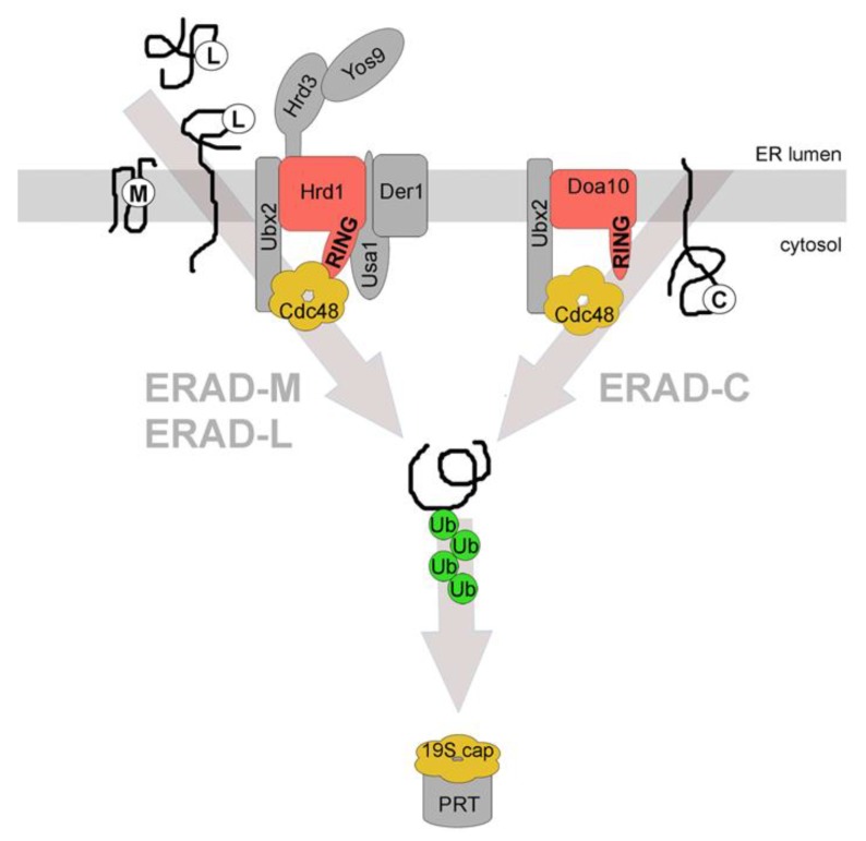
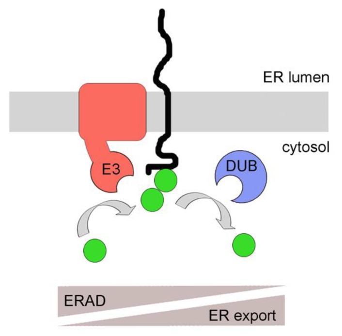
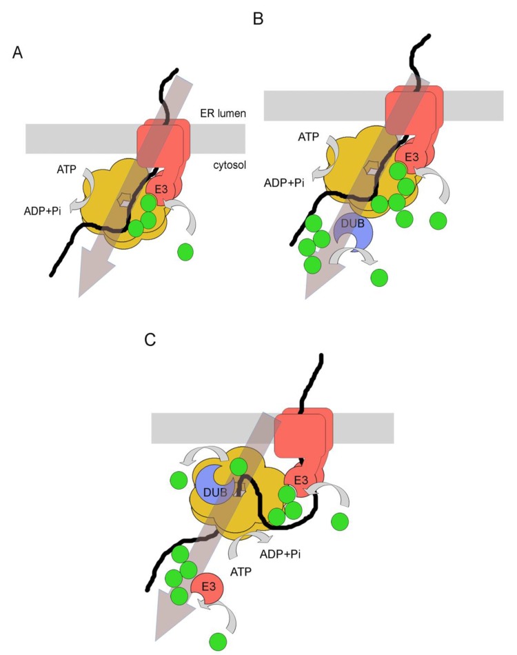
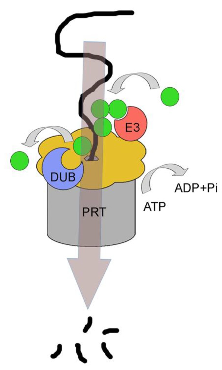
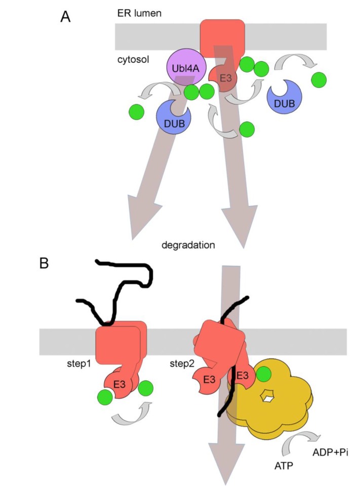
Quality control of protein folding inside the endoplasmic reticulum (ER) includes chaperone-mediated assistance in folding and the selective targeting of terminally misfolded species to a pathway called ER-associated protein degradation, or simply ERAD. Once selected for ERAD, substrates will be transported (back) into the cytosol, a step called retrotranslocation. Although still ill defined, retrotranslocation likely involves a protein conducting channel that is in part formed by specific membrane-embedded E3 ubiquitin ligases. Early during retrotranslocation, reversible self-ubiquitination of these ligases is thought to aid in initiation of substrate transfer across the membrane. Once being at least partially exposed to the cytosol, substrates will become ubiquitinated on the cytosolic side of the ER membrane by the same E3 ubiquitin ligases. Ubiquitin on substrates was originally thought to be a permanent modification that (1) promotes late steps of retrotranslocation by recruiting the energy-providing ATPase Cdc48p/p97 via binding to its associated adaptor proteins and that (2) serves to target substrates to the proteasome. Recently it became evident, however, that the poly-ubiquitin chains (PUCs) on ERAD substrates are often subject to extensive remodeling, or processing, at several stages during ERAD. This review recapitulates the current knowledge and recent findings about PUC processing on ERAD substrates and ubiquitination of ERAD machinery components and discusses their functional consequences.


Figures

Figure 1
Figure 1
Distinct endoplasmic reticulum-associated degradation (ERAD) pathways. Three main ERAD pathways in yeast are classified based on substrates and the components that are involved in their degradation. ERAD-L degrades membrane integrated or soluble proteins with misfolded domains in the ER lumen, marked with (L). All depicted constituents of the Hrd1-complex are required for the efficient degradation of these substrates. ERAD-M degrades membrane integrated proteins with misfolded regions in their transmembrane domain(s), marked with (M). Proteins of this class are degraded via the Hrd1-complex but do not require Usa1p and Der1p for efficient degradation. ERAD-C degrades membrane integrated proteins with misfolded domains in the cytoplasm, marked with (C). These proteins are degraded via the Doa10-complex. All ERAD pathways require cytosolic Cdc48p for substrate retrotranslocation and extraction from the ER membrane. The substrate is ubiquitinated by the E3 ligases during or after retrotranslocation and is targeted to the proteasome (PRT) for degradation. Cdc48p and the 19S cap of the proteasome have structural and functional similarities. The classification for the different ERAD pathways also exists in mammalian cells albeit it is less stringent. RING = RING domain of the E3 ligases that are shown in red. The associated constituents of the individual complexes are shown in grey. Ub = ubiquitin. See text for details.
Figure 2
Figure 2
A model for poly-ubiquitin chain (PUC) processing as a mechanism for coupling ER protein quality control with ERAD. Membrane proteins with exposed cytosolic domains are ubiquitinated by E3 ligases. Due to topological confinement of membrane proteins and membrane integrated E3 ligases to the same bilayer, ubiquitination might occur frequently, even on correctly folded proteins. DUBs can remove ubiquitins, favoring ER export of correctly folded species. Reoccurring ubiquitination due to prolonged ER retention as a result of misfolding (in the cytosol or ER lumen) will favor the assembly of PUCs to induce targeting to the proteasome, thereby favoring ERAD. A similar mechanism might occur for soluble proteins prior to their post-translational translocation across the ER membrane, a pathway called prERAD. See text for details. E3 = E3 ligase. DUB = deubiquitinase. Filled green circles: ubiquitin.
Figure 3
Figure 3
Models for PUC processing as a mechanism for protein retrotranslocation. (A) Ubiquitination of the substrate on the cytosolic side of the ER membrane by an E3 ligase promotes binding to the hexameric Cdc48p, via its ubiquitin-binding cofactors Npl4p and Ufd1p. ATP hydrolysis by Cdc48p is thought to promote the complete retrotranslocation and substrate extraction from the ER membrane. See text for details. (B) In the mammalian system, the p97-associated DUB Ataxin-3 acts downstream of protein retrotranslocation and might promote the removal of ubiquitins from longer PUCs. This is thought to be required for the generation of PUCs with optimal binding affinities to proteasomal shuttle factors or to the proteasome itself. See text for details. (C) The p97-associated DUB YOD1 is thought to act upstream of protein retrotranslocation. Its activity was suggested to be needed for the complete removal of PUCs to allow passage of the substrate through the pore of p97. ATP hydrolysis would then couple protein retrotranslocation with protein unfolding. Subsequently, reubiquitination by an E3 ligase would allow substrate targeting to the proteasome. See text for details.
Figure 4
Figure 4
The 19S cap of the proteasome has functional and structural similarities with Cdc48p/p97. Like Cdc48p/p97, the hexameric 19S cap of the proteasome binds simultaneously to E3 ligases and to DUBs, suggesting that PUCs are processed at this stage, immediately before substrate degradation. This could be needed for the transfer of the substrate from the periphery of the proteasomal lid through its central pore and further into the proteolytic chamber of the proteasome. Immediately before passage through the pore of the l9S cap, structural data suggest that all PUCs are to be removed. See text for details.
Figure 5
Figure 5
Models for ubiquitination and deubiquitination of ERAD machinery components as a mechanism to regulate ERAD. (A) Regulation through degradation. In the mammalian system, ubiquitination of the ERAD component Ubl4A by the E3 ligase gp78 leads to the degradation of the associated shuttling chaperone Bag6, thereby reducing efficient substrate targeting to the proteasome. Ubl4A ubiquitination is reversed by the DUB Usp13. A similar mechanism involves the self-ubiquitination of Hrd1p in yeast in absence of Hrd3p and Usa1p, leading to effective degradation of the E3 ligase. No DUB is currently known in this system. See text for details. (B) Regulation through conformational changes. Binding of a substrate to Hrd1p in the ER lumen induces oligomerization and self-ubiquitination of the E3 ligase (step 1). Subsequently, ATP hydrolysis by bound Cdc48p would result in conformational changes in the ATPase, which will consequently result in conformational changes in Hrd1p oligomers. This, in turn, would push the substrate through a postulated channel formed by Hrd1p oligomers (together with associated components like Der1p) until it is partially exposed to the cytosol (step 2). See text for details.

References

    1. Rapoport T.A. Protein translocation across the eukaryotic endoplasmic reticulum and bacterial plasma membranes. Nature. 2007;450:663–669. doi: 10.1038/nature06384. - DOI - PubMed
    1. Sommer T., Jentsch S. A protein translocation defect linked to ubiquitin conjugation at the endoplasmic reticulum. Nature. 1993;365:176–179. doi: 10.1038/365176a0. - DOI - PubMed
    1. Hiller M.M., Finger A., Schweiger M., Wolf D.H. ER degradation of a misfolded luminal protein by the cytosolic ubiquitin-proteasome pathway. Science. 1996;273:1725–1728. doi: 10.1126/science.273.5282.1725. - DOI - PubMed
    1. Werner E.D., Brodsky J.L., McCracken A.A. Proteasome-dependent endoplasmic reticulum-associated protein degradation: An unconventional route to a familiar fate. Proc. Natl. Acad. Sci. USA. 1996;93:13797–13801. doi: 10.1073/pnas.93.24.13797. - DOI - PMC - PubMed
    1. Schulman B.A., Harper J.W. Ubiquitin-like protein activation by E1 enzymes: The apex for downstream signalling pathways. Nat. Rev. Mol. Cell. Biol. 2009;10:319–331. doi: 10.1038/nrm2673. - DOI - PMC - PubMed

LinkOut - more resources