. 2014 Aug 7:5:4623.
doi: 10.1038/ncomms5623.

Single-molecule force spectroscopy reveals force-enhanced binding of calcium ions by gelsolin

Affiliations

Single-molecule force spectroscopy reveals force-enhanced binding of calcium ions by gelsolin

Chunmei Lv et al. Nat Commun. .

Abstract

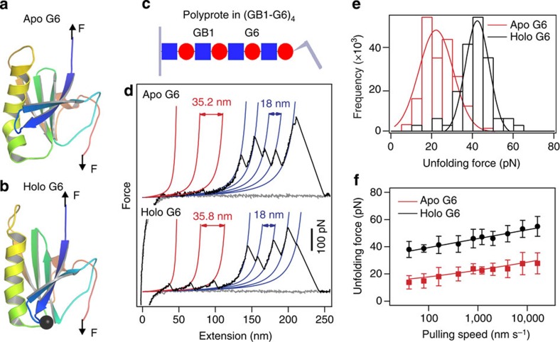
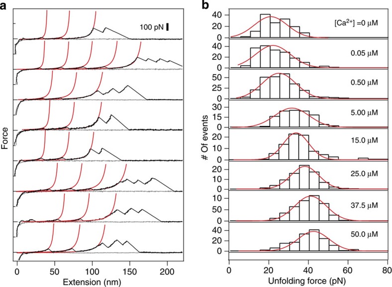
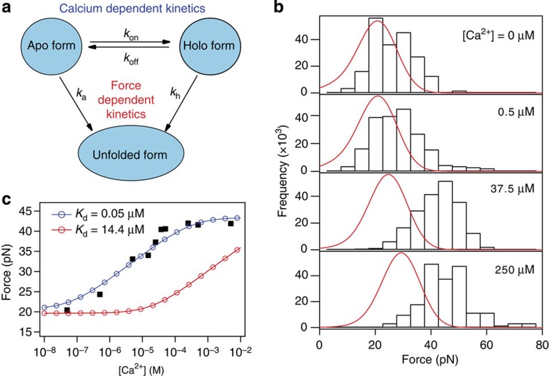
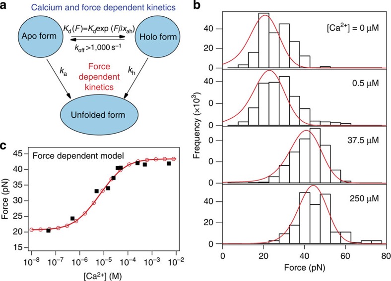
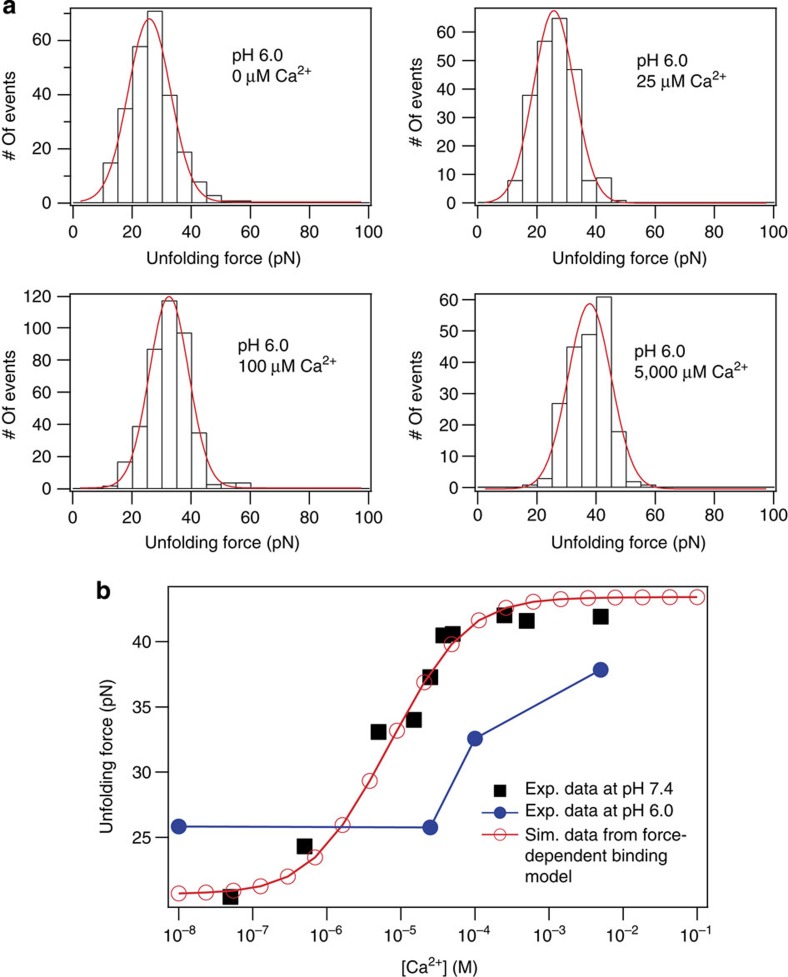
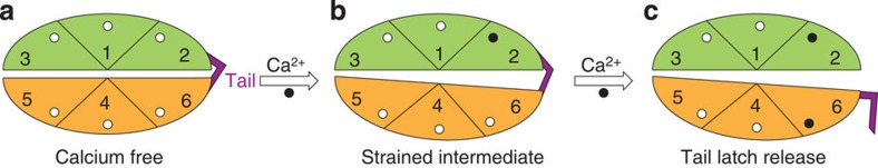
Force is increasingly recognized as an important element in controlling biological processes. Forces can deform native protein conformations leading to protein-specific effects. Protein-protein binding affinities may be decreased, or novel protein-protein interaction sites may be revealed, on mechanically stressing one or more components. Here we demonstrate that the calcium-binding affinity of the sixth domain of the actin-binding protein gelsolin (G6) can be enhanced by mechanical force. Our kinetic model suggests that the calcium-binding affinity of G6 increases exponentially with force, up to the point of G6 unfolding. This implies that gelsolin may be activated at lower calcium ion levels when subjected to tensile forces. The demonstration that cation-protein binding affinities can be force-dependent provides a new understanding of the complex behaviour of cation-regulated proteins in stressful cellular environments, such as those found in the cytoskeleton-rich leading edge and at cell adhesions.


Figures

Figure 1
Figure 1. Mechanical properties of G6 probed by single-molecule AFM.
The structure of G6 in the absence of Ca2+ (PDB ID: 3FFN) (a) and in the presence of Ca2+ (shown as black sphere), (PDB ID: 1P8X) (b). Upon Ca2+ binding, the central long helix is straightened. The schematic representations are shown in rainbow colours from blue (N termini) to red (C termini). The C-terminal latch sequence is not shown because it becomes unstructured in holo G6 and was not evident in the crystal structures of gelsolin-actin complexes (Supplementary Fig. 1). However, the C-terminal helix sequence was included in the polyprotein (GB1–G6)4 for single-molecule AFM experiments. The arrows indicate the direction of applied force is from the N to C terminus of G6. (c) A schematic of the hetero-polyprotein (GB1–G6)4 used for single-molecule force spectroscopy experiments. Red circles and blue squares represent G6 and GB1 domains, respectively. (d) Typical set of approaching (grey line) and retraction traces for mechanical unfolding of calcium-free apo or calcium-bound holo forms of (GB1–G6)4 at a pulling speed of 400 nm s−1. WLC fitting of consecutive unfolding events (red and blue lines) identifies that all saw-tooth-like peaks result from the unfolding of G6 and GB1 domains with contour length increments of 35 nm and 18 nm, respectively. The unfolding force for G6 increases from ~20 to ~40 pN upon saturation with Ca2+ (at ~50 μM) while the unfolding force for GB1 remains the same. (e) Unfolding force histograms of apo and holo G6. (f) Pulling speed dependency of apo and holo G6. The unfolding force increases with increasing pulling speed. Error bars represent standard deviations. The experimental data can be numerically fitted using a two-state unfolding model (solid lines). The unfolding rate constant at zero force (k0) and the unfolding distance (Δxu) are 0.09 s−1 and 1.73 nm for apo G6, and 5.0 × 10⁻⁵ s−1 and 1.60 nm for holo G6, respectively.
Figure 2
Figure 2. Mechanical unfolding of G6 at different [Ca2+].
(a) Representative force-extension profiles for unfolding of (GB1–G6)4 in the presence of various [Ca2+] at a pulling speed of 400 nm s−1. The [Ca2+] at which the traces were measured are shown on the same row in b. Red lines correspond to the WLC fitting to the G6 unfolding events. (b) The unfolding forces histograms at different [Ca2+]. The solid lines are Gaussian fittings of these data. The unfolding forces of G6 show unimodal distributions at all Ca2+ concentrations and the widths of the distributions remain constant. This indicates that the calcium-free and calcium-bound conformations are in fast equilibrium upon stretching.
Figure 3
Figure 3. A force-independent binding model.
(a) The kinetic model that considers rapid dynamics between the calcium-free apo and the calcium-bound holo conformations. The unfolding of both the apo and holo forms are force-dependent processes and the interconversion between the apo and holo forms depends on the calcium concentration and is force independent. (b) This kinetic model fails to reproduce the unfolding force histograms at various calcium concentrations. Black bars correspond to the experimentally obtained unfolding force histograms and red lines correspond to the numeric fitting results using the kinetic model depicted in a The χ2 for the fittings to the histograms from top to bottom are 0.093, 0.096, 0.308 and 0.290, respectively. (c) This kinetic model cannot describe the unfolding forces of G6 at different calcium concentrations. Red circles correspond to the numerically calculated data using a Kd of 14.4 μM, as determined from ITC measurements. Blue circles correspond to the numerically calculated data using a Kd of 0.05 μM that best ‘fits’ the experimental data. However, the slope of the unfolding force versus [Ca2+] relationship cannot be fully reproduced. The χ2 for numerically calculated data using Kds of 14.4 and 0.05 μM are 14.0 and 0.33, respectively.
Figure 4
Figure 4. A force-dependent binding model.
(a) In this new kinetic model, both the unfolding kinetics of the calcium-free apo and the calcium-bound holo forms and their interconversion are force-dependent processes. xah is a parameter describing the dependency of dissociation constant on the applied force. We set koff>1,000 s−1 to reflect the fast interconversion between apo and holo forms. (b) The unfolding force histograms at various calcium concentrations (black bars) can be numerically fitted using the Kd obtained from ITC and an xah of −0.7 nm based on the ‘force-dependent binding model’ (red lines). The negative xah indicates that force decreases the dissociation constant and makes the holo form more stable. The χ2 for the fittings to the histograms from top to bottom are 0.093, 0.059, 0.036 and 0.029, respectively. (c) This kinetic model can also describe the unfolding forces of G6 at different calcium concentrations. Red circles and black squares correspond to the numerically calculated data and the experimentally obtained data, respectively. The χ2 for the fitting is 0.22.
Figure 5
Figure 5. The change of the unfolding forces for G6 at different [Ca2+] at pH 6.
(a) The unfolding force histograms of G6 at different [Ca2+], pH 6 (pulling speed: 400 nm s−1). (b) The dependency of the unfolding forces of G6 on the concentrations of calcium ions at pH 7.4 and 6.0. Exp., experimental; Sim., Simulation.
Figure 6
Figure 6. Cartoon highlighting the initial activation stages of gelsolin.
(a) Six-domain gelsolin contains a homologous calcium-binding site in each domain (white circles) and a C-terminal tail that interacts with domain 2 to lock the inactive conformation (coloured in purple). (b) Occupation of any of these sites by a calcium ion will begin the activation process that drives conformational reorganization and concomitantly creates inter-domain strain, leading to the C-terminal tail under tension. Here we show a calcium ion occupying the site on G2. Greater strain may be induced through filling more of the calcium-binding sites. (c) The resulting inter-domain strain will increase the affinity of G6 for calcium, resulting in calcium ion binding to G6 and the release of the tail latch, which in turn exposes the F-actin-binding site on G2.

Similar articles

Cited by

References

    1. Silacci P. et al. Gelsolin superfamily proteins: key regulators of cellular functions. Cell. Mol. Life Sci. 61, 2614–2623 (2004). - PMC - PubMed
    1. Nag S., Larsson M., Robinson R. C. & Burtnick L. D. Gelsolin: the tail of a molecular gymnast. Cytoskeleton (Hoboken) 70, 360–384 (2013). - PubMed
    1. Sun H. Q., Yamamoto M., Mejillano M. & Yin H. L. Gelsolin, a multifunctional actin regulatory protein. J. Biol. Chem. 274, 33179–33182 (1999). - PubMed
    1. Kiselar J. G., Janmey P. A., Almo S. C. & Chance M. R. Visualizing the Ca2+-dependent activation of gelsolin by using synchrotron footprinting. Proc. Natl Acad. Sci. USA 100, 3942–3947 (2003). - PMC - PubMed
    1. Ashish et al. Global structure changes associated with Ca2+ activation of full-length human plasma gelsolin. J. Biol. Chem. 282, 25884–25892 (2007). - PubMed

Publication types