Review
. 2014 Aug;20(8):822-32.
doi: 10.1038/nm.3642.

Lung regeneration: mechanisms, applications and emerging stem cell populations

Affiliations
Review

Lung regeneration: mechanisms, applications and emerging stem cell populations

Darrell N Kotton et al. Nat Med. 2014 Aug.

Abstract

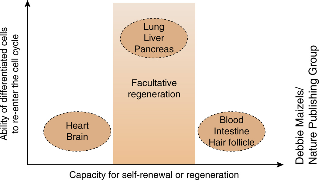
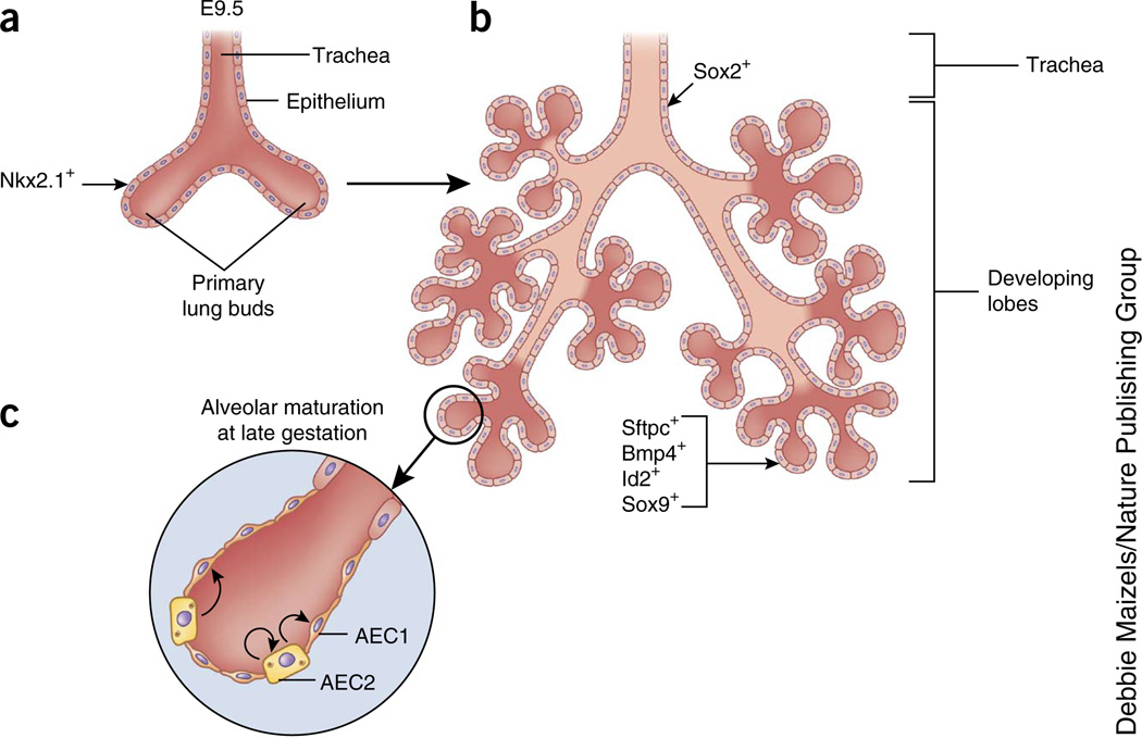
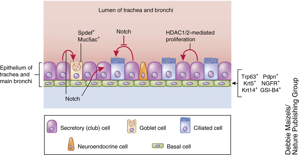
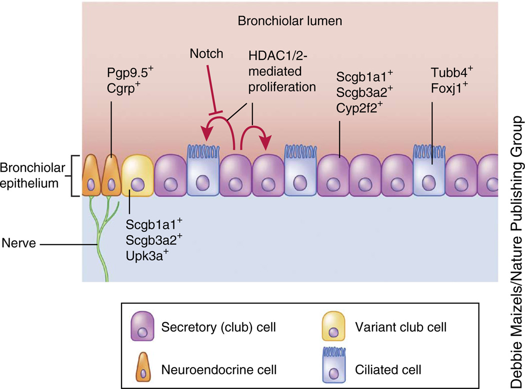
Recent studies have shown that the respiratory system has an extensive ability to respond to injury and regenerate lost or damaged cells. The unperturbed adult lung is remarkably quiescent, but after insult or injury progenitor populations can be activated or remaining cells can re-enter the cell cycle. Techniques including cell-lineage tracing and transcriptome analysis have provided novel and exciting insights into how the lungs and trachea regenerate in response to injury and have allowed the identification of pathways important in lung development and regeneration. These studies are now informing approaches for modulating the pathways that may promote endogenous regeneration as well as the generation of exogenous lung cell lineages from pluripotent stem cells. The emerging advances, highlighted in this Review, are providing new techniques and assays for basic mechanistic studies as well as generating new model systems for human disease and strategies for cell replacement.


Figures

Figure 1
Figure 1
Relationship between the regenerative capacity of different tissues and the existence of resident tissue-specific stem cells. Tissues such as the hematopoietic system and the intestine undergo rapid turnover assisted by well-documented stem cell lineages. Other tissues, such as the lung, can respond robustly after injury to replace lost cells but are normally quiescent in the adult. A third group of tissues, including the heart and brain, does not regenerate well after injury and generally forms scar tissue. Differentiated cells in tissues that undergo rapid turnover do not exhibit the robust ability to re-enter the cell cycle, whereas facultative regenerative tissues, such as the lung, do.
Figure 2
Figure 2
Cell lineages in early lung development in mouse. a. At E9.5 in mouse, the trachea and two primary lung buds are formed. b. These buds subsequently branch into lobes, which develop into alveoli. Proximal and distal developing epithelia of the developing lungs express different markers. At this early stage, Id2+ cells can generate proximal and distal epithelia. c. Toward late gestation, AEC1s and AEC2s are formed.
Figure 3
Figure 3
Stem cell and differentiated epithelia ineages in the trachea and main stem bronch of the lung. The trachea and most proxima airways of the rodent and human lung are lined with multiple epithelial lineages. Basal cells are located in this region and can generate secretory and ciliated cell lineages. Cell signaling pathways such as Notch are crucial for differentiation of basal cells and also suppress the ciliated epithelial–cell fate. HDAC1 and HDAC2 (HDAC1/2) are essential for secretory epithelial regeneration. Red arrows indicate cells that have been shown by lineage-tracing techniques to generate the indicated lineages after injury or during homeostasis.
Figure 4
Figure 4
Stem cell and differentiated epithelial lineages in the bronchiolar airways of the lung. The rodent lung contains a bronchiolar region that acks basal cells but is lined with a simple cuboidal epithelium. Notch suppresses the ciliated epithelial cell fate in this region of the airways. HDAC1 and HDAC2 (HDAC1/2) are essential for regeneration of secretory epithelial lineages, such as club cells. Red arrows indicate cells that have been shown by lineage-tracing techniques to generate the ndicated lineages after injury or during homeostasis.
Figure 5
Figure 5
Progenitor cell populations and their differentiated progeny in the lung alveolus. The alveolar epithelium consists of AEC1s and AEC2s. Lineage-tracing techniques indicate that AEC2s can generate AEC1s during homeostasis and after injury (red arrow). Generation of alveolar epithelium by other cells, such as Sftpc+Scgb1a1+ (putative BASCs) and Itgb4+cells, has yet to be supported by lineage tracing (black arrows). One study also suggests that basal cells can generate alveolar epithelium, although additional lineage tracing is required to support this hypothesis. Red arrows indicate cells that have been shown by lineage-tracing techniques to generate the indicated lineages after injury or during homeostasis. In the case of the SftpcItgb4+ population, direct lineage tracing has not yet been reported. Wnt signaling is thought to be important in regulating alveolar epithelial homeostasis after injury, and Wnt signaling and Gata6 are important for BASC expansion and differentiation. a6, Itga6; β4, Itgb4.

Similar articles

Cited by

References

    1. Beers MF, Morrisey EE. The three R’s of lung health and disease: repair, remodeling, and regeneration. J. Clin. Invest. 2011;121:2065–2073. - PMC - PubMed
    1. Wansleeben C, Barkauskas CE, Rock JR, Hogan BL. Stem cells of the adult lung: their development and role in homeostasis, regeneration, and disease. Wiley Interdiscip. Rev. Dev. Biol. 2013;2:131–148. - PubMed
    1. Zaret KS, Grompe M. Generation and regeneration of cells of the liver and pancreas. Science. 2008;322:1490–1494. - PMC - PubMed
    1. Peng T, et al. Coordination of heart and lung co-development by a multipotent cardiopulmonary progenitor. Nature. 2013;500:589–592. - PMC - PubMed
    1. Cardoso WV, Lu J. Regulation of early lung morphogenesis: questions, facts and controversies. Development. 2006;133:1611–1624. - PubMed