. 2014 Aug 6;34(32):10624-34.
doi: 10.1523/JNEUROSCI.0335-14.2014.

Hydrogen peroxide increases GABAA receptor-mediated tonic current in hippocampal neurons

Affiliations

Hydrogen peroxide increases GABAA receptor-mediated tonic current in hippocampal neurons

Antonello Penna et al. J Neurosci. .

Abstract

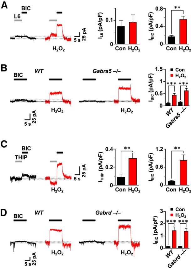
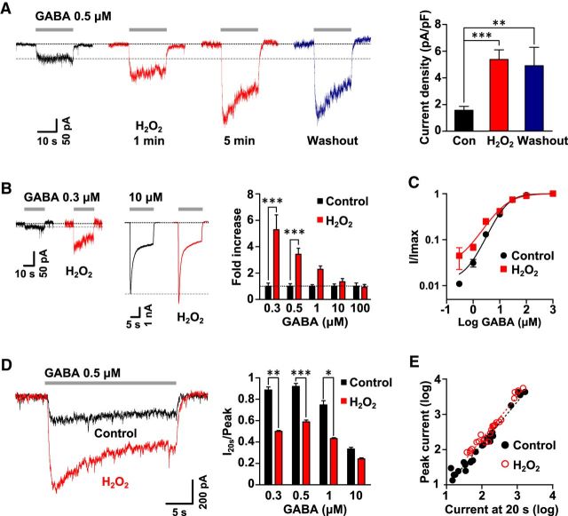
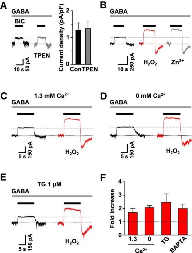
Hydrogen peroxide (H2O2), a key reactive oxygen species, is produced at low levels during normal cellular metabolism and at higher concentrations under pathological conditions such as ischemia-reperfusion injury. The mechanisms by which H2O2 contributes to physiological and pathological processes in the brain remain poorly understood. Inhibitory GABA type A (GABAA) receptors critically regulate brain function by generating tonic and synaptic currents; however, it remains unknown whether H2O2 directly modulates GABAA receptor function. Here, we performed patch-clamp recordings, together with pharmacological and genetic approaches, to investigate the effects of H2O2 on GABAA receptor-mediated tonic and synaptic currents recorded in cultured mouse hippocampal neurons and CA1 pyramidal neurons in hippocampal slices. We found that H2O2 caused a dramatic increase in tonic current, whereas synaptic currents were unaffected. This increase in tonic current resulted from an extracellular oxidative reaction, which increased the potency of GABA, but only when GABAA receptors were activated by low concentrations of GABA. Oxygen-glucose deprivation, which produces high endogenous levels of H2O2, similarly increased the tonic current. These results suggest that GABAA receptor-mediated tonic current, which is potentiated by H2O2, might contribute to H2O2-induced brain dysfunction.

Keywords: GABAA receptor; hippocampus; hydrogen peroxide; ischemia-reperfusion; mouse; tonic current.


Figures

Figure 1.
Figure 1.
H2O2 increased tonic GABA currents in hippocampal neurons. A, Representative traces and summarized data showing that the tonic current revealed by BIC (100 μm) was increased after 5 min of H2O2 (200 μm, n = 21) and this increase persisted for at least 5 min after washout (n = 6). B, Representative trace and summarized data showing the time-dependent effects of H2O2 (200 μm) on tonic current in the presence of a low concentration of GABA (0.5 μm, n = 17–23). C, Representative traces and summarized data demonstrate that tonic current was persistently increased even after 20 min of washout in the presence of GABA (0.5 μm, n = 5–7). One-way ANOVA: F(2,47) = 16.1, for A; F(4,102) = 6.7 for B, F(4,24) = 0.4; and p = 0.4 for C; Newman–Keuls post hoc test, **p < 0.01 and ***p < 0.001. D, Left, Representative traces showing the increase in tonic current induced by a low-concentration (10 μm) and a high-concentration (1000 μm) of H2O2 in the presence of GABA (0.5 μm). Right: Quantified data demonstrating the dose-dependent enhancing effects of H2O2 on tonic current (n = 4 for each concentration). Two-way ANOVA, effect of H2O2: F(1,30) = 32.0, p < 0.0001; effect of concentration: F(4,30) = 9.2, p < 0.0001; effect of interaction: F(4,30) = 9.2, p < 0.0001; Bonferroni post hoc test, **p < 0.01, ***p < 0.001. Con, Control (for here and in subsequent figures).
Figure 2.
Figure 2.
H2O2 had no effect on synaptic GABA currents. A, Representative recordings of GABAA receptor-mediated mIPSCs recorded at −60 mV before (left) and after (right) 5 min of H2O2 (200 μm) treatment from the same neuron. B, Traces were averaged from 475 (control) and 527 (H2O2) individual mIPSCs. C, Cumulative amplitude (left) and frequency (right) distributions of mIPSCs showing that both the amplitude and the frequency were not affected by H2O2; p = 0.24 for the amplitude, p = 0.06 for the frequency, Kolmogorov–Smirnov test.
Figure 3.
Figure 3.
H2O2 increased tonic GABA currents in hippocampal slices. Representative traces (A) and summarized data (B) showing the increase in tonic current after 10 min of H2O2 (1 mm) treatment (n = 6); *p = 0.02, Student's unpaired t test.
Figure 4.
Figure 4.
α5GABAA and δGABAA receptors were not required for the H2O2-dependent increase in current. A, Left, Representative traces showing the tonic current revealed by a selective inverse agonist of α5GABAA receptors (L6, 20 nm) and BIC (100 μm). The L6-sensitive tonic current was unaffected, whereas BIC-sensitive current was greatly increased by H2O2. Right, Quantification of L6- and BIC-sensitive tonic current, before and after H2O2 (n = 6); **p = 0.002, Student's paired t test. B, Left, Representative traces showing the tonic current revealed by BIC (100 μm) in neurons from WT and Gabra5−/− mice. Right, Quantification of the tonic current before and after H2O2 (n = 10 for WT, n = 9 for Gabra5−/−). C, Left, Representative traces showing the current activated by δGABAA receptor-preferring agonist THIP (0.5 μm) and the tonic current revealed by BIC (100 μm). Right, Quantification of THIP-activated inward current (n = 6) and the tonic current revealed by BIC (n = 5) before and after H2O2; **p = 0.007 for THIP, **p = 0.008 for BIC, Student's paired t test. D, Left, Representative traces showing the tonic current revealed by BIC in neurons from WT and Gabrd−/− mice. Right, Quantification of the tonic current before and after H2O2 (n = 9 for each genotype).
Figure 5.
Figure 5.
H2O2 increased the potency of low concentrations of GABA without modifying the desensitization properties. A, Left, Representative traces showing the time-dependent effects of H2O2 (200 μm) on currents evoked by a low concentration of GABA (0.5 μm). Right, Summarized data (n = 4–13). One-way ANOVA: F(2,27) = 11.8; Newman–Keuls post hoc test; **p < 0.01, ***p < 0.001. B, Left, Representative traces showing the effects of H2O2 (200 μm) on the currents evoked by 0.3 and 10 μm GABA. Right, Summarized data showing the fold increase in currents evoked by different concentrations of GABA by H2O2 (200 μm; n = 5–13). Two-way ANOVA, effect of H2O2: F(1,58) = 33.2, p < 0.0001; effect of concentration: F(4,58) = 7.6, p < 0.0001; effect of interaction: F(4,58) = 7.6, p < 0.0001; Bonferroni post hoc test, ***p < 0.001. C, Concentration-response plots in the absence and presence of H2O2 fitted with Hill equation (n = 8–11). D, Left, Representative traces showing currents evoked by GABA (0.5 μm) before and after H2O2 (200 μm). Right, Summarized data showing the effects of H2O2 on desensitization of currents evoked by increasing concentrations of GABA. H2O2 decreased the ratio of current at 20 s to peak current only at low GABA concentrations but not at 10 μm (n = 4–9). Two-way ANOVA, effect of H2O2: F(1,38) = 33.5, p < 0.0001; effect of concentration: F(3,38) = 18.0, p < 0.0001; effect of interaction: F(3,38) = 1.8, p = 0.16; Bonferroni post hoc test, *p < 0.05, **p < 0.01, ***p < 0.001. E, Correlation between the current at 20 s and peak current, before and after H2O2 (200 μm). The linear regression fit generated similar slopes between control and H2O2 treatment (n = 23 for each condition).
Figure 6.
Figure 6.
H2O2 did not modify the voltage sensitivity of the tonic GABA current. A, The I–V relationship of tonic current before and after H2O2 treatment was studied using a voltage step protocol for holding potentials between −80 and +20 mV that had a duration of 4 s (n = 5–6). B, Summarized data showing similar increase in tonic current by H2O2 at all membrane potentials (n = 5). One-way ANOVA: F(4,20) = 0.17, p = 0.95.
Figure 7.
Figure 7.
Changes in the concentration of Zn2+ and Ca2+ did not modify the effects of H2O2. A, Left, Representative traces showing the effects of TPEN (10 μm) on the tonic current. Right, Quantification of the increase in tonic current before and after TPEN (n = 4). B, Representative traces showing the effects of Zn2+ (10 μm) on tonic current after being potentiated by H2O2 (200 μm). Similar results were obtained from another three cells. C–E, Representative traces showing the increase in tonic current by H2O2 (200 μm) in the presence of normal extracellular Ca2+ (C), in the absence of extracellular Ca2+ (D), or when intracellular Ca2+ was depleted with thapsigargin (TG, 1 μm; E). F, Summarized data for experiments in C–E (n = 5–9 for each condition). One-way ANOVA: F(3,20) = 0.7, p = 0.55.
Figure 8.
Figure 8.
Extracellular oxidative reaction mediated the effects of H2O2. A, Addition of the antioxidant GSH (1 mm) to the pipette solution did not modify the increase in tonic current by H2O2 (200 μm). n = 6 for each group, **p = 0.008, Student's unpaired t test. B, C, Addition of the antioxidants GSH (1 mm) and DTT (1 mm) to the extracellular solution abolished the increase in tonic current by H2O2 (200 μm); n = 4. D, DFO (100 μm) prevented the increase in tonic current by H2O2 (200 μm); n = 6.
Figure 9.
Figure 9.
OGD treatment increased tonic current through an oxidative reaction. A, Reperfusion after OGD increased tonic current; n = 9, **p = 0.006, Student's paired t test. B, C, The increase in tonic current by reperfusion was abolished and a decrease after reperfusion was observed for neurons treated with the antioxidant GSH (1 mm; B) or pretreated for 30 min with NADPH oxidase inhibitor DPI (10 μm; C). Student's paired t test; n = 7, *p = 0.03 for B; n = 7, ***p < 0.001 for C.

Similar articles

Cited by

References

    1. Abramov AY, Canevari L, Duchen MR. β-Amyloid peptides induce mitochondrial dysfunction and oxidative stress in astrocytes and death of neurons through activation of NADPH oxidase. J Neurosci. 2004;24:565–575. doi: 10.1523/JNEUROSCI.4042-03.2004. - DOI - PMC - PubMed
    1. Abramov AY, Scorziello A, Duchen MR. Three distinct mechanisms generate oxygen free radicals in neurons and contribute to cell death during anoxia and reoxygenation. J Neurosci. 2007;27:1129–1138. doi: 10.1523/JNEUROSCI.4468-06.2007. - DOI - PMC - PubMed
    1. Allen CL, Bayraktutan U. Oxidative stress and its role in the pathogenesis of ischaemic stroke. Int J Stroke. 2009;4:461–470. doi: 10.1111/j.1747-4949.2009.00387.x. - DOI - PubMed
    1. Andersen JK. Oxidative stress in neurodegeneration: cause or consequence? Nat Med. 2004;10:S18–S25. doi: 10.1038/nrn1434. - DOI - PubMed
    1. Bianchi MT, Macdonald RL. Agonist trapping by GABAA receptor channels. J Neurosci. 2001;21:9083–9091. - PMC - PubMed

Publication types

MeSH terms

LinkOut - more resources