. 2014 Oct 3;289(40):27665-76.
doi: 10.1074/jbc.M114.570341. Epub 2014 Aug 6.

Characterization of the influence of mediator complex in HIV-1 transcription

Affiliations

Characterization of the influence of mediator complex in HIV-1 transcription

Alba Ruiz et al. J Biol Chem. .

Abstract

HIV-1 exploits multiple host proteins during infection. siRNA-based screenings have identified new proteins implicated in different pathways of the viral cycle that participate in a broad range of cellular functions. The human Mediator complex (MED) is composed of 28 elements and represents a fundamental component of the transcription machinery, interacting with the RNA polymerase II enzyme and regulating its ability to express genes. Here, we provide an evaluation of the MED activity on HIV replication. Knockdown of 9 out of 28 human MED proteins significantly impaired viral replication without affecting cell viability, including MED6, MED7, MED11, MED14, MED21, MED26, MED27, MED28, and MED30. Impairment of viral replication by MED subunits was at a post-integration step. Inhibition of early HIV transcripts was observed by siRNA-mediated knockdown of MED6, MED7, MED11, MED14, and MED28, specifically affecting the transcription of the nascent viral mRNA transactivation-responsive element. In addition, MED14 and MED30 were shown to have special relevance during the formation of unspliced viral transcripts (p < 0.0005). Knockdown of the selected MED factors compromised HIV transcription induced by Tat, with the strongest inhibitory effect shown by siMED6 and siMED14 cells. Co-immunoprecipitation experiments suggested physical interaction between MED14 and HIV-1 Tat protein. A better understanding of the mechanisms and factors controlling HIV-1 transcription is key to addressing the development of new strategies required to inhibit HIV replication or reactivate HIV-1 from the latent reservoirs.

Keywords: Host Factor; Human Immunodeficiency Virus (HIV); RNA Polymerase II; RNA Silencing; RNAP II; Tat; Transcription; Viral Transcription.


Figures

FIGURE 1.
FIGURE 1.
Expression profile of Mediator complex mRNA in different cell types and tissues. mRNA expression of each Mediator gene was quantified by qPCR in samples of RNA extracted from TZM-bl cells, MT-4 cells, PBMC, monocytes, MDM, and commercial lymph node, adipose, small intestine, and thymus RNA. Normalized expression of different mRNAs was calculated using GAPDH quantification as reference. Results from gene expression assays were uploaded to the GEPAS software to obtain a cluster image representing gene expression from low (blue) to high (red) expression and functional profiling of the Mediator complex. The mean of two MED expression profile assays is shown.
FIGURE 2.
FIGURE 2.
Inhibition of HIV-1 replication after siRNA-mediated interference of Mediator complex subunits. A, mRNA knockdown efficiency in TZM-bl cells was quantified by qPCR 48 h post-transfection. mRNA expression of each MED gene was normalized to a sample treated without siRNA (Mock). Results from one of three independent experiments are shown. B, TZM-bl cells were transfected with the indicated siRNA and infected 48 h later with HIV-1 NL4-3 strain for 72 h. A nontargeting (NT) siRNA pool and a siRNA targeting CD4 were used as controls. Infection was measured by quantifying expression of β-gal reporter and validated with a dose-response effect of the reverse transcription inhibitor AZT (white bars). Viability of transfected cells was monitored by the MTT method and validated with a DMSO dose-response effect (black bars). Means ± S.D. of at least three independent experiments are shown. *, p < 0.05. C, different siRNA targeting MED14 confirmed the antiviral effect at subtoxic concentrations. HeLa TZM-bl cells were transfected with four different siRNAs targeting MED14 at different molarities, and mRNA levels were quantified by qPCR 48 h later. Transfected cells were then infected with HIV-1 NL4-3 strain, and 72 h later infection was quantified or cells were assayed for siRNA-derived toxicity by the MTT assay. A result of one representative experiment of three is shown.
FIGURE 3.
FIGURE 3.
MED14 and MED27 impaired HIV-1 replication in MDM. A, mRNA knockdown efficiency in MDM cells was quantified by qPCR 72 h post-transfection. mRNA expression of each MED gene was normalized to a sample treated without siRNA (Mock). Means ± S.D. of three independent experiments are shown. B, MDM were transfected with the indicated siRNA and infected (INF) 72 h later with vesicular stomatitis virus-pseudotyped NL4-3-GFP virus. Infection was measured as the percentage of GFP-positive cells in siRNA-treated macrophages and expressed as the percentage to mock-treated cells. AZT was used as a control (white bars). In parallel, cell viability was assayed in uninfected (UN) siRNA-treated macrophages by counting live cells using flow cytometry (black bars).Means ± S.D. of three independent donors are shown. RALT, raltegravir. *, p < 0.05.
FIGURE 4.
FIGURE 4.
Selected Mediator subunits have a role in HIV-1 cycle at the early and late transcription level. Integrated proviral DNA (gray bars), TAR RNA (white bars), and tat/rev/nef late viral transcripts (black bars) were compared by qPCR in siRNA-treated TZM-bl cells infected for 24 h (for integrated proviral DNA) or 48 h (for TAR and late viral transcripts) with NL4-3 HIV-1 strain. Values are expressed as a percentage of the control cells treated with an NT siRNA. The RT inhibitor AZT, the integrase inhibitor raltegravir (RALT), and a siRNA targeting ZNRD1 gene (ZNRD1) were used as controls. UN, uninfected. Means ± S.D. of three independent experiments are shown. *, p < 0.05; **, p < 0.005; ***, p < 0.0005.
FIGURE 5.
FIGURE 5.
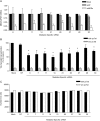
Mediator complex has a role in Tat-driven expression of HIV-1 LTR promoter. A, indicated siRNAs were transfected into HeLa TZM-bl cells. After 48 h, an expression vector of HIV-1 Tat was also transfected. After an additional 24 h, RNA samples were obtained to quantify MED mRNA levels by qPCR. Expression levels were normalized to the Mock-transfected sample, and an NT siRNA was used as a control. Mean ± S.D. of three independent experiments are shown. B, β-gal assay was performed in samples treated as in A, either transfected with a Tat-expressing plasmid (black bars) or transfected in the absence of Tat plasmid (white bars). Values were normalized to the mock control. NT siRNA was used as control. Means ± S.D. of three independent experiments are shown. *, p < 0.05. C, samples treated as in A were recovered, and viability was monitored by counting viable cells by flow cytometry in Tat-plasmid transfected cells (black bars) or pcTat-untransfected cells (white bars).
FIGURE 6.
FIGURE 6.
Co-immunoprecipitation of HIV-1 Tat with MED14. A, lysates from untransfected (Mock) HEK293T cells, transfected with a FLAG-Tat expression plasmid (FLAG-Tat) or transfected with an untagged Tat expression plasmid (pcTat), were subjected to immunoprecipitation (IP) with anti-FLAG antibodies attached to agarose (lanes 1–3) or-Sepharose alone (lane 4). Immunoprecipitates were then blotted with an anti-MED14 antibody or anti-Tat antibody. WCL (lanes 5–7) were blotted with anti-MED14 or anti-Hsp90 antibodies as a control. acryl, acrylamide. B, lysates used in A were subjected to immunoprecipitation with anti-MED14 antibody attached to Sepharose (lanes 1–4). Immunoprecipitates were probed by immunoblotting analysis with an anti-Tat antibody or anti-MED14 antibody. WCL were subjected to SDS-PAGE and blotted with anti-Tat or anti-β-actin antibodies (lanes 5–7). One representative experiment of three is shown. C, benzonase treatment was performed before the immunoprecipitation following same procedure as in A. DNA from untreated or treated protein lysates was extracted and quantified by qPCR. Values were relativized and expressed as 2∧ (−RNase P amplification cycle). D, HEK293T cells, previously transfected with a FLAG-Tat expression plasmid (FLAG-Tat) or mock-transfected, were then retrotransfected with siRNA targeting mRNA from indicated MED subunits. mRNA expression of each MED gene was normalized to a sample treated without siRNA. Results from one of two independent experiments are shown. E and F, cell lysates treated as described in D were then subjected to immunoprecipitation with anti-FLAG antibodies attached to agarose (beads). Immunoprecipitates were then blotted with an anti-MED14 antibody or anti-Tat antibody. G, WCL from E and F were blotted with anti-MED14, anti-Hsp90, anti-Tat, or anti-β-actin antibodies as a control.

Similar articles

Cited by

References

    1. Ballana E., Esté J. A. (2013) Insights from host genomics into HIV infection and disease: identification of host targets for drug development. Antiviral Res. 100, 473–786 - PubMed
    1. Van Lint C., Bouchat S., Marcello A. (2013) HIV-1 transcription and latency: an update. Retrovirology 10, 67. - PMC - PubMed
    1. Ruelas D. S., Greene W. C. (2013) An integrated overview of HIV-1 latency. Cell 155, 519–529 - PMC - PubMed
    1. Marcello A., Zoppé M., Giacca M. (2001) Multiple modes of transcriptional regulation by the HIV-1 Tat transactivator. IUBMB Life 51, 175–181 - PubMed
    1. Kao S. Y., Calman A. F., Luciw P. A., Peterlin B. M. (1987) Anti-termination of transcription within the long terminal repeat of HIV-1 by tat gene product. Nature 330, 489–493 - PubMed

Publication types