. 2014 Sep 26;289(39):27264-27277.
doi: 10.1074/jbc.M114.578435. Epub 2014 Aug 6.

Conformational analysis of the Streptococcus pneumoniae hyaluronate lyase and characterization of its hyaluronan-specific carbohydrate-binding module

Affiliations

Conformational analysis of the Streptococcus pneumoniae hyaluronate lyase and characterization of its hyaluronan-specific carbohydrate-binding module

Michael D L Suits et al. J Biol Chem. .

Abstract

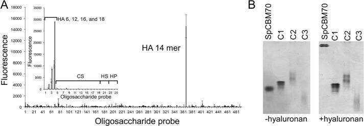
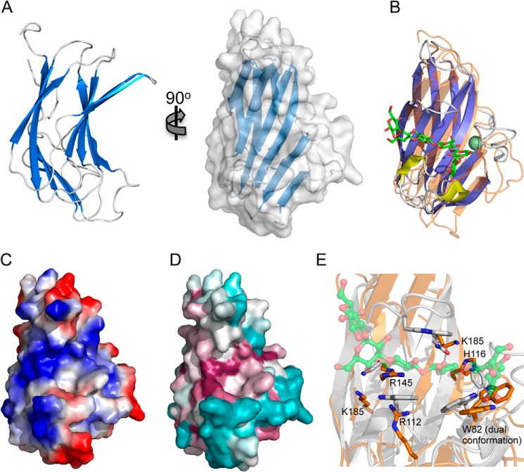
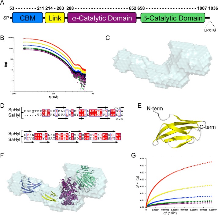
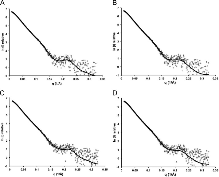
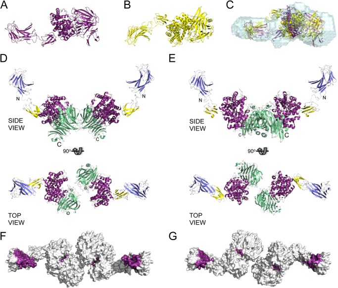
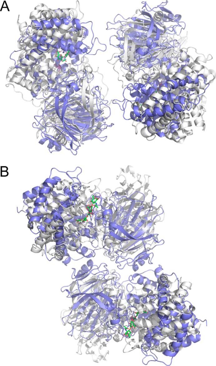
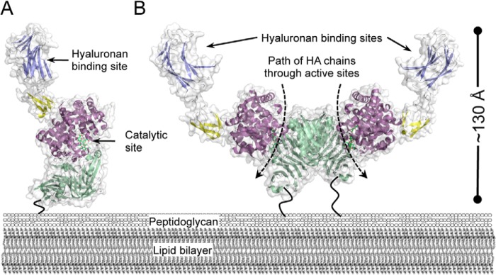
For a subset of pathogenic microorganisms, including Streptococcus pneumoniae, the recognition and degradation of host hyaluronan contributes to bacterial spreading through the extracellular matrix and enhancing access to host cell surfaces. The hyaluronate lyase (Hyl) presented on the surface of S. pneumoniae performs this role. Using glycan microarray screening, affinity electrophoresis, and isothermal titration calorimetry we show that the N-terminal module of Hyl is a hyaluronan-specific carbohydrate-binding module (CBM) and the founding member of CBM family 70. The 1.2 Å resolution x-ray crystal structure of CBM70 revealed it to have a β-sandwich fold, similar to other CBMs. The electrostatic properties of the binding site, which was identified by site-directed mutagenesis, are distinct from other CBMs and complementary to its acidic ligand, hyaluronan. Dynamic light scattering and solution small angle x-ray scattering revealed the full-length Hyl protein to exist as a monomer/dimer mixture in solution. Through a detailed analysis of the small angle x-ray scattering data, we report the pseudoatomic solution structures of the monomer and dimer forms of the full-length multimodular Hyl.

Keywords: Carbohydrate; Carbohydrate Processing; Carbohydrate-binding Protein; Crystallography; Glycobiology; Hyaluronan; Hyaluronate Lyase; Small Angle X-ray Scattering (SAXS); Streptococcus.


Figures

FIGURE 1.
FIGURE 1.
Analyses of the glycan binding properties of SpCBM70. A, microarray analysis of SpCBM70 binding specificity. The screening glycan microarray of 492 lipid-linked probes, NGLs, and glycosylceramides (>370 mammalian type) is the Glycosciences Array Set 32–39 described previously (21) (supplemental Table 1). Binding signals are expressed as numerical scores, means of fluorescence intensities of duplicate values recorded at 5 fmol oligosaccharide probe/spot. The error bars represent half of the difference between the two values. The inset shows microarray analyses with 25 lipid-linked GAG oligosaccharide probes. Numerical scores of the binding signals are means of duplicate spots (with error bars) of oligosaccharide probes printed at 2 (light gray bars) and 7 (dark gray bars) fmol/spot on nitrocellulose-coated glass slides. HA, CS, HS, and HP, hyaluronan, chondroitin sulfate, heparin sulfate, and heparin fragments, respectively. B, native polyacrylamide gel electrophoresis analysis of SpCBM70 in the absence of hyaluronan (−hyaluronan) and with hyaluronan present in the gel at a final concentration of 0.045% (w/v) (+hyaluronan). C1, C2, and C3, the migration of three non-interacting control proteins.
FIGURE 2.
FIGURE 2.
The structure of SpCBM70. A, schematic representation of the 1.2 Å resolution structure of SpCBM70 shown from two angles and with the solvent-accessible surface shown in transparent gray (right). B, a structural overlap of SpCBM70 (transparent orange) with TmCBM4–2 (blue/yellow) from Thermotoga maritima (PDB entry 1GUI). The β-1,3-glucohexaose molecule bound to TmCBM4–2 is shown as green sticks, and the bound calcium is shown as a green sphere. C, the solvent-accessible surface of SpCBM70 (shown from the same angle as in B) colored by electrostatic potential. D, the solvent-accessible surface of SpCBM70 (shown from the same angle as in B) colored by conservation of the residues among similar modules from Streptococci (generated using ConSurf (54)). E, detailed view of the overlap of the putative SpCBM70 hyaluronan binding site (orange) with the β-1,3-glucohexaose binding site in TmCBM4-2 (gray). Residues targeted for mutagenesis in SpCBM70 are shown as sticks and labeled.
FIGURE 3.
FIGURE 3.
Modular properties and initial SAXS analysis of full-length Hyl. A, schematic of the modular architecture of Hyl. The α- and β-catalytic domains constitute the PL8 domain. B, SAXS scattering profiles of Hyl, protein concentrations: 10.80 (red), 5.40 (yellow), 2.70 (blue), 1.50 (green), 1.35 (black), and 1.00 mg/ml (purple). C, DAMAVER averaged shape of 10 independent DAMMIF-generated envelopes of Hyl using the data collected on the 1.0 mg/ml sample. D, alignment of the linker module from Hyl with the similar module from the Streptococcus agalactiae Hyl (SaHyl). E, Phyre2-generated model using the structure of the SaHyl linker domain as a template. F, the composite modules of Hyl (CBM (blue), linker (yellow), α-catalytic domain (purple), and β-catalytic domain (green)) manually placed into the DAMMIF envelope. G, Porod-Debye plots of the SAXS data for Hyl. A, q4·I(q) versus q4 plot for Hyl at 10.80 (red), 5.40 (yellow), 2.70 (blue), 2.4 (green), 1.35 (black), and 1.00 mg/ml (purple).
FIGURE 4.
FIGURE 4.
SASREFMX and OLIGOMER fits of monomer dimer populations. A, SASREF7MX fit for best model of population A; B, OLIGOMER fit for best model of population A; C, SASREF7MX fit for best model of population B; D, OLIGOMER fit for best model of population B. Models were generated with SASREF7MX for Hyl at 1.00 mg/ml.
FIGURE 5.
FIGURE 5.
SAXS-based rigid body modeling of Hyl as an oligomeric mixture. A, model of the Hyl monomer present in population A generated by SASREF7MX rigid body modeling of Hyl as a three-module protein in a monomer/dimer equilibrium. B, as in A but the Hyl monomer present in population B. C, monomers of population A (purple) and population B (yellow) overlapped with the DAMMIF-generated envelopes of Hyl (1.0 mg/ml) using SUPCOMB. The monomers in A and B are shown in the same orientation as they are in the overlapped view in C. D and E, dimer structures of populations A and B, respectively, shown from similar side and top views. The modules are colored as follows: CBM (blue), linker (yellow), α-catalytic domain (purple), and β-catalytic domain (green). The N and C termini are labeled for reference. F and G, top views of the dimer structures of populations A and B, respectively, as solvent-accessible surface areas with the binding sites of the CBMs colored purple and the active site of the PL8 domain colored purple and with a bound substrate (green sticks; modeled from the structure of the PL8 domain in complex with a 4-mer fragment of hyaluronan; PDB entry 1LXK).
FIGURE 6.
FIGURE 6.
Comparison of the SAXS-based solution dimers of Hys (CBM and linker not shown for clarity) with dimers of the PL8 domain generated by crystallographic packing in the C2 crystal form (PDB entry 1BRV). A, the dimer of the PL8 domain in population A (gray) overlapped with one crystallographic dimer of the PL8 domain (blue). B, the dimer of the PL8 domain in population B (gray) overlapped with the alternate crystallographic dimer of the PL8 domain (blue).
FIGURE 7.
FIGURE 7.
Modeled surface presentation of Hyl. Models of the Hyl monomer (A) and dimer (B) as they may be presented on the surface of the bacterium. Only the structures from population A of the SAXS-generated modules are shown because the two populations are very similar in overall organization. The dashed arrows represent the path of processive cleavage of hyaluronan (HA) chains through the PL8 domain active sites. The dark, bending lines represent the unmodeled C-terminal portions of Hyl that tether the protein to the peptidoglycan through a sortase-mediated process.

References

    1. Jiang D., Liang J., Noble P. W. (2011) Hyaluronan as an immune regulator in human diseases. Physiol. Rev. 91, 221–264 - PMC - PubMed
    1. Hava D. L., Camilli A. (2002) Large-scale identification of serotype 4 Streptococcus pneumoniae virulence factors. Mol. Microbiol. 45, 1389–1406 - PMC - PubMed
    1. Rapport M. M., Linker A., Meyer K. (1951) The hydrolysis of hyaluronic acid by pneumococcal hyaluronidase. J. Biol. Chem. 192, 283–291 - PubMed
    1. Thompson R. T., Moses F. E. (1948) Elaboration of hyaluronidase by pneumococci isolated from bacteremic pneumococcic pneumonia patients. J. Clin. Invest. 27, 558. - PubMed
    1. Musser J. M., Mattingly S. J., Quentin R., Goudeau A., Selander R. K. (1989) Identification of a high-virulence clone of type III Streptococcus agalactiae (group B Streptococcus) causing invasive neonatal disease. Proc. Natl. Acad. Sci. U.S.A. 86, 4731–4735 - PMC - PubMed

Publication types

MeSH terms

Associated data