. 2014 Aug 6;6(248):248ra106.
doi: 10.1126/scitranslmed.3008930.

Immunoglobulin class-switched B cells form an active immune axis between CNS and periphery in multiple sclerosis

Affiliations

Immunoglobulin class-switched B cells form an active immune axis between CNS and periphery in multiple sclerosis

Arumugam Palanichamy et al. Sci Transl Med. .

Abstract

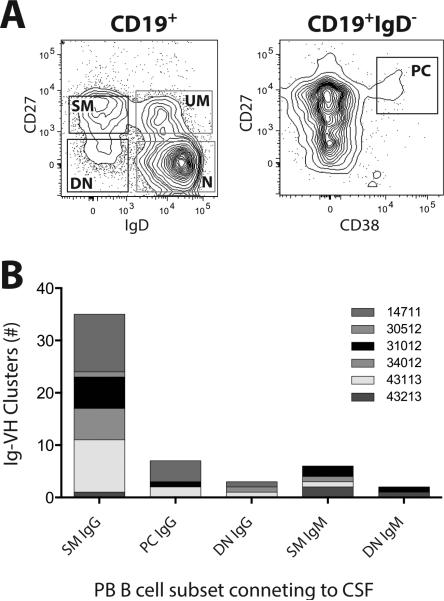
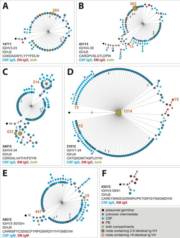
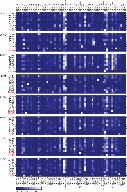
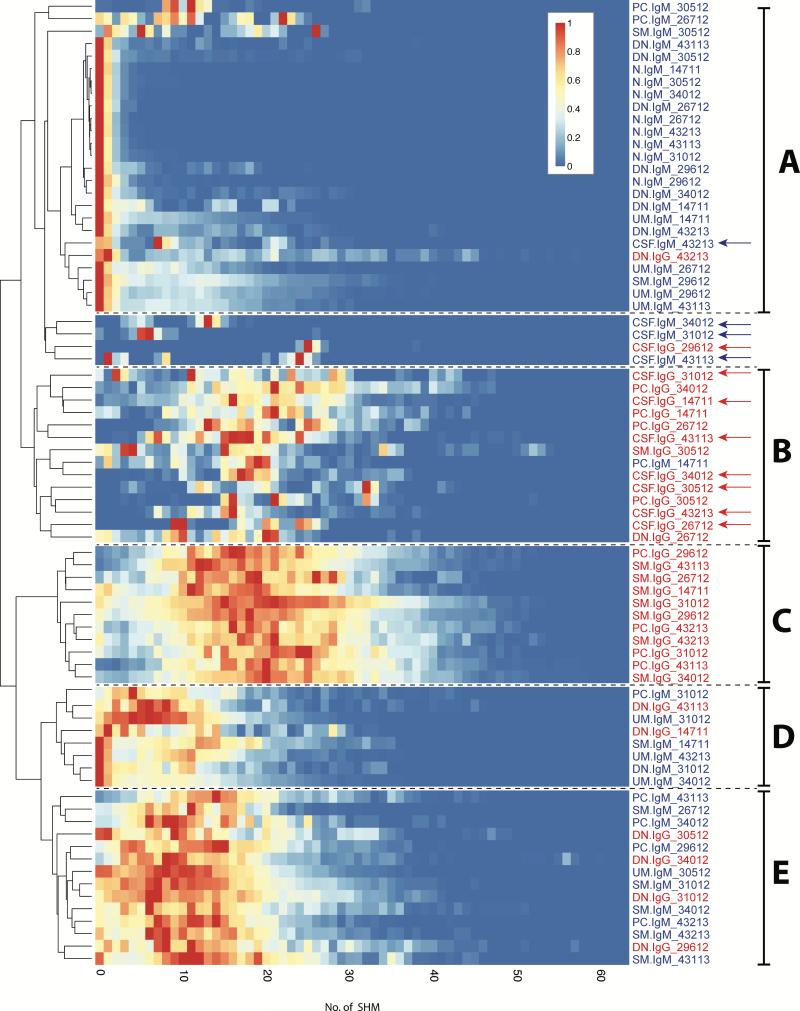
In multiple sclerosis (MS), lymphocyte--in particular B cell--transit between the central nervous system (CNS) and periphery may contribute to the maintenance of active disease. Clonally related B cells exist in the cerebrospinal fluid (CSF) and peripheral blood (PB) of MS patients; however, it remains unclear which subpopulations of the highly diverse peripheral B cell compartment share antigen specificity with intrathecal B cell repertoires and whether their antigen stimulation occurs on both sides of the blood-brain barrier. To address these questions, we combined flow cytometric sorting of PB B cell subsets with deep immune repertoire sequencing of CSF and PB B cells. Immunoglobulin (IgM and IgG) heavy chain variable (VH) region repertoires of five PB B cell subsets from MS patients were compared with their CSF Ig-VH transcriptomes. In six of eight patients, we identified peripheral CD27(+)IgD(-) memory B cells, CD27(hi)CD38(hi) plasma cells/plasmablasts, or CD27(-)IgD(-) B cells that had an immune connection to the CNS compartment. Pinpointing Ig class-switched B cells as key component of the immune axis thought to contribute to ongoing MS disease activity strengthens the rationale of current B cell-targeting therapeutic strategies and may lead to more targeted approaches.


Figures

Figure 1
Figure 1. Peripheral blood B cell subsets connecting to the CNS compartment
A: Representative FACS plots (patient 34012) illustrating PB B cell subset identification and sorting strategy; plasmablasts/plasma cells (PC) are CD27hiCD38hi gated on CD19+IgD cells; switched memory (SM), unswitched memory (UM), double negative (DN), and naïve (N) B cells were sorted based on the presence or absence of CD27 or IgD gated on CD19+ cells. Sorting gates are indicated in the FACS plots, see Figure S7 for single color histograms. PB B cell subsets with connections to the CSF are in black gates, those not connecting to the CSF are in gray gates. B: PB B cell clusters connecting to the CNS; each column represents the sum of Ig-VH clusters with contributions from CSF and the indicated PB B cell subset.
Figure 2
Figure 2. Affinity-maturation of B cells connecting CSF and PB
Shown are representative lineages (A to F) of clonally related Ig-VH found in both compartments. Lineages were calculated using IgTree (see Methods) and visualized in Cytoscape using the proprietary Organic layout option. Numbers between nodes represent numbers of nucleotide mutations; connections without numbers represent a single nucleotide mutation. Round nodes without rim encompass a single Ig-VH sequence; round nodes with black rims contain 2 to 9 identical Ig-VH, those with orange rims contain >9 identical Ig-VH sequences (orange number next to respective nodes). Closest putative germline sequences represent the root of each lineage (black nodes). Gray nodes are lineage intermediates that were not identified as Ig-VH transcript and were calculated by IgTree. Shown are also IGHV and IGHJ used per lineage and representative H-CDR3. Alignments of representative Ig-VH are shown in Figure S5.
Figure 3
Figure 3. IGHV usage in PB B cell subsets and CSF
Shown are heatmaps representing relative usage of IGHV germline segments by IgM-VH or IgG-VH from the indicated PB B cell subsets or CSF for each patient studied (Table S6). To generate heatmaps, IGHV usage values were normalized to the most frequently used IGHV per sample and Ig isotype, which were set as 1. The resulting heatmaps show the most frequently used IGHV in white and the least frequently used IGHV in dark blue (see lower left corner for scale). Notably, IGHV usage is generally limited in CSF (red type) suggesting selective recruitment or survival of B cells in the CSF compartment. IGHV usage of CSF IgG-VH in patient 14711 is more balanced and similar peripheral B cell repertoires; this patient had signs of active disease 2 weeks prior to lumbar puncture (Table 1).
Figure 4
Figure 4. Dendogram and heatmaps of PB B cell and CSF Ig-VH SHM profiles
Shown are heatmaps of Ig-VH SHM profiles; each row represents a PB B cell subset or CSF as indicated by row titles on the right. IgM are in blue type, IgG are in red type; CSF samples are indicated by arrows. Within each subset or CSF sample, SHM profiles were scaled to the highest peak set at 1.0 and colors assigned such that a count of 0 non-redundant sequences with a certain umber of SHM resulted in blue color and the highest number of sequences with an indicated number of SHM was labeled red; the coloring scale is shown in the upper right. PB B cells subsets or CSF samples clustering together based on SHM profile-similarity are indicated by letters on the right (see also Results). A: Group of subsets characterized by predominantly low SHM, including all naïve B cells and nearly all IgM+ DN B cells. B and C: Groups of subsets characterized by extensive SHM, and containing all IgG-expressing SM B cells and plasma cells, and the majority of CSF samples. D and E: Subsets characterized by SHM lower than B and C, but higher than A; these groups include mainly IgM-expressing subsets and IgG-expressing DN B cells. The data used to generate these heatmaps is shown in Table S8.

Comment in

References

    1. Bar-Or A, Calabresi PA, Arnold D, Markowitz C, Shafer S, Kasper LH, Waubant E, Gazda S, Fox RJ, Panzara M, Sarkar N, Agarwal S, Smith CH. Rituximab in relapsing-remitting multiple sclerosis: a 72-week, open-label, phase I trial. Annals of neurology. 2008;63:395–400. - PubMed
    1. Hauser SL, Waubant E, Arnold DL, Vollmer T, Antel J, Fox RJ, Bar-Or A, Panzara M, Sarkar N, Agarwal S, Langer-Gould A, Smith CH, Group HT. B-cell depletion with rituximab in relapsing-remitting multiple sclerosis. N Engl J Med. 2008;358:676–688. - PubMed
    1. Kappos L, Li D, Calabresi PA, O'Connor P, Bar-Or A, Barkhof F, Yin M, Leppert D, Glanzman R, Tinbergen J, Hauser SL. Ocrelizumab in relapsingremitting multiple sclerosis: a phase 2, randomised, placebo-controlled, multicentre trial. Lancet. 2011;378:1779–1787. - PubMed
    1. Lovato L, Willis SN, Rodig SJ, Caron T, Almendinger SE, Howell OW, Reynolds R, O'Connor KC, Hafler DA. Related B cell clones populate the meninges and parenchyma of patients with multiple sclerosis. Brain : a journal of neurology. 2011;134:534–541. - PMC - PubMed
    1. Obermeier B, Lovato L, Mentele R, Bruck W, Forne I, Imhof A, Lottspeich F, Turk KW, Willis SN, Wekerle H, Hohlfeld R, Hafler DA, O'Connor KC, Dornmair K. Related B cell clones that populate the CSF and CNS of patients with multiple sclerosis produce CSF immunoglobulin. Journal of neuroimmunology. 2011;233:245–248. - PMC - PubMed

Publication types

MeSH terms

Substances

Associated data