. 2014 Oct;88(20):11696-712.
doi: 10.1128/JVI.01433-14. Epub 2014 Aug 6.

The 3' untranslated region of Pea Enation Mosaic Virus contains two T-shaped, ribosome-binding, cap-independent translation enhancers

Affiliations

The 3' untranslated region of Pea Enation Mosaic Virus contains two T-shaped, ribosome-binding, cap-independent translation enhancers

Feng Gao et al. J Virol. 2014 Oct.

Abstract

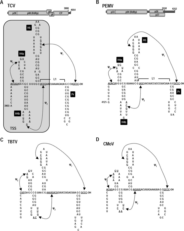
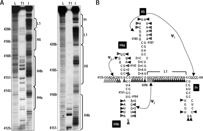
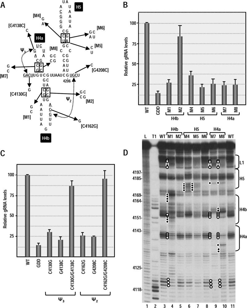
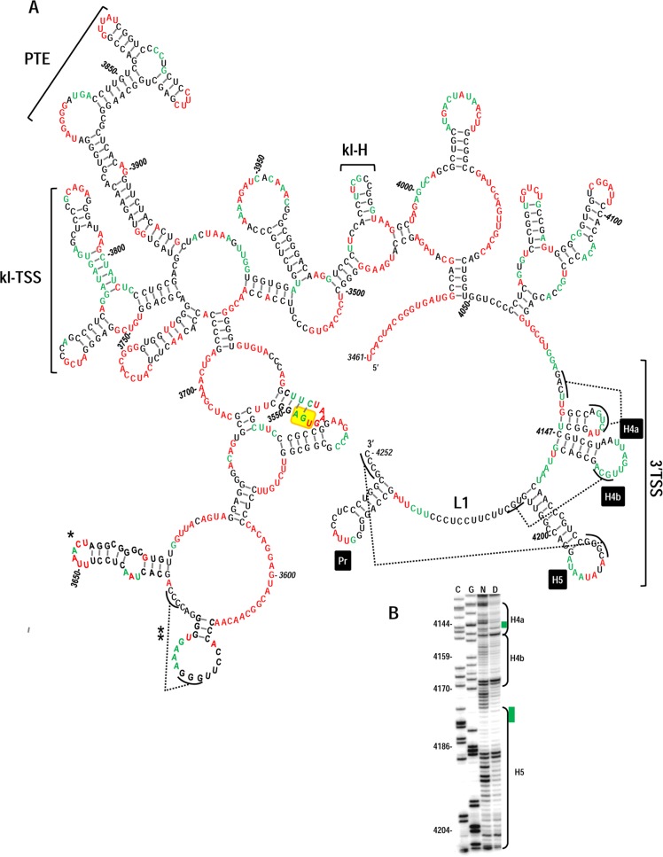
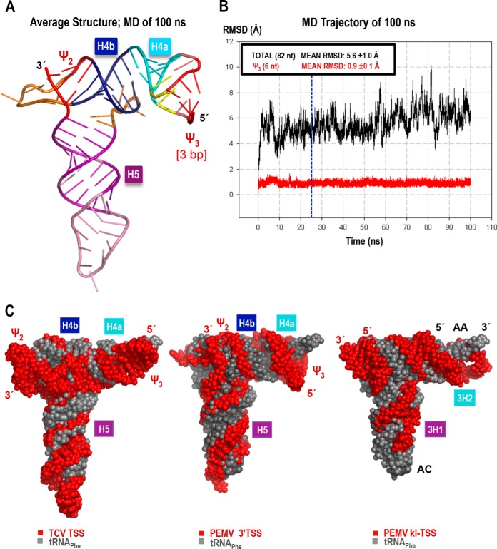
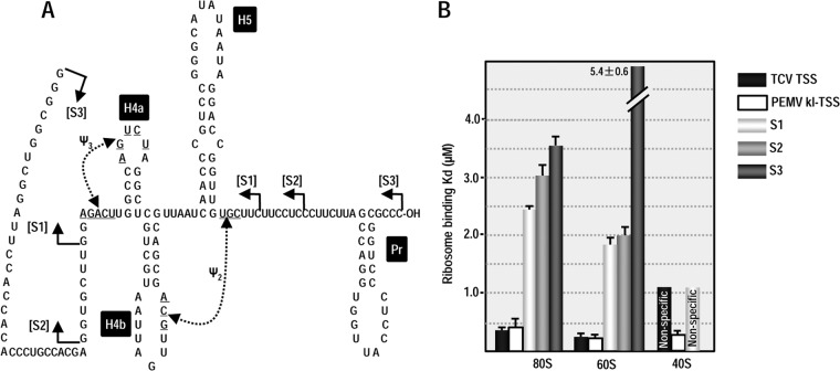
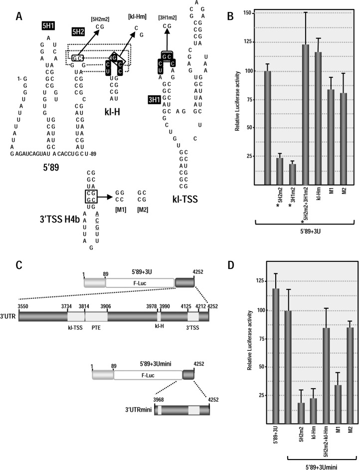
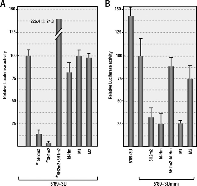
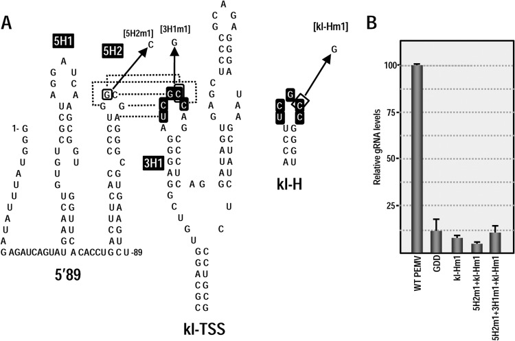
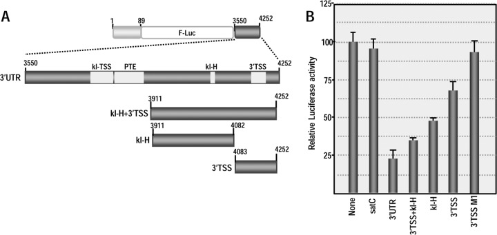
Many plant viruses without 5' caps or 3' poly(A) tails contain 3' proximal, cap-independent translation enhancers (3'CITEs) that bind to ribosomal subunits or translation factors thought to assist in ribosome recruitment. Most 3'CITEs participate in a long-distance kissing-loop interaction with a 5' proximal hairpin to deliver ribosomal subunits to the 5' end for translation initiation. Pea Enation Mosaic Virus (PEMV) contains two adjacent 3'CITEs in the center of its 703-nucleotide 3' untranslated region (3'UTR), the ribosome-binding, kissing-loop T-shaped structure (kl-TSS) and eukaryotic translation initiation factor 4E-binding Panicum mosaic virus-like translation enhance (PTE). We now report that PEMV contains a third, independent 3'CITE located near the 3' terminus. This 3'CITE is composed of three hairpins and two pseudoknots, similar to the TSS 3'CITE of the carmovirus Turnip crinkle virus (TCV). As with the TCV TSS, the PEMV 3'TSS is predicted to fold into a T-shaped structure that binds to 80S ribosomes and 60S ribosomal subunits. A small hairpin (kl-H) upstream of the 3'TSS contains an apical loop capable of forming a kissing-loop interaction with a 5' proximal hairpin and is critical for the accumulation of full-length PEMV in protoplasts. Although the kl-H and 3'TSS are dispensable for the translation of a reporter construct containing the complete PEMV 3'UTR in vitro, deleting the normally required kl-TSS and PTE 3'CITEs and placing the kl-H and 3'TSS proximal to the reporter termination codon restores translation to near wild-type levels. This suggests that PEMV requires three 3'CITEs for proper translation and that additional translation enhancers may have been missed if reporter constructs were used in 3'CITE identification. Importance: The rapid life cycle of viruses requires efficient translation of viral-encoded proteins. Many plant RNA viruses contain 3' cap-independent translation enhancers (3'CITEs) to effectively compete with ongoing host translation. Since only single 3'CITEs have been identified for the vast majority of individual viruses, it is widely accepted that this is sufficient for a virus's translational needs. Pea Enation Mosaic Virus possesses a ribosome-binding 3'CITE that can connect to the 5' end through an RNA-RNA interaction and an adjacent eukaryotic translation initiation factor 4E-binding 3'CITE. We report the identification of a third 3'CITE that binds weakly to ribosomes and requires an upstream hairpin to form a bridge between the 3' and 5' ends. Although both ribosome-binding 3'CITEs are critical for virus accumulation in vivo, only the CITE closest to the termination codon of a reporter open reading frame is active, suggesting that artificial constructs used for 3'CITE identification may underestimate the number of CITEs that participate in translation.


Figures

FIG 1
FIG 1
Conserved structural features at the 3′ ends of TCV and three umbraviruses. (A) Top, genome organization of the carmovirus TCV. p28 is a replication-required protein, and its readthrough product p88 is the viral RdRp. p8 and p9 movement proteins are expressed from one sgRNA, and the capsid protein (CP) is expressed from a second sgRNA. Bottom, secondary and tertiary structure elements at the 3′ terminus of TCV. All 15 carmoviruses contain Pr, H5, and ψ1. Most carmoviruses contain H4b and fewer contain both H4b and ψ2 (63). Only TCV, CCFV, and JINRV contain all hairpins and pseudoknots shown (23). The boxed, shaded region denotes that hairpins H4a, H4b, H5, and pseudoknots ψ2 and ψ3 fold into a 3-D T-shaped structure (TSS). (B) Top, Genome organization of PEMV. p33 is a putative replication-required protein, and its −1 ribosomal frameshift protein is the RdRp. p26 and p27 are movement required proteins expressed from the single sgRNA. Bottom, possible structural organization at the 3′ terminus of PEMV based on the structures found in TCV. (C and D) Phylogenetically conserved elements at the 3′ end of TBTV (C) and CMoV (D).
FIG 2
FIG 2
In-line probing of the 3′ end of PEMV. (A) Susceptibility of residues at the 3′ end of PEMV to in-line cleavage. The 170-nt 3′ terminal fragment was radiolabeled at the 5′ end and incubated at 25°C for 14 h, followed by denaturing gel electrophoresis. The locations of different putative 3′ elements are indicated to the right of each autoradiogram. L, OH treated ladder; T1, partial RNase T1 digest to denote location of guanylates; I, in-line cleavage of the fragment. The intensity of each band is proportional to the flexibility of the residue at that location. (B) Susceptibility of residues in the putative structure of PEMV 3′ region to in-line cleavage. Darker triangles denote stronger cleavage.
FIG 3
FIG 3
Effect of alterations in 3′ elements on PEMV accumulation in protoplasts. (A) Single and compensatory mutations generated in the 3′ hairpins and pseudoknots that may comprise a TSS. The names of the alterations are indicated in brackets. (B) Relative levels of full-length WT PEMV and PEMV containing alterations in 3′ hairpins accumulating in Arabidopsis protoplasts at 24 h after inoculation. Standard deviations from three independent experiments are shown. GDD, PEMV nonreplicating control with an altered GDD RdRp active-site motif. (C) Relative levels of full-length PEMV accumulating in Arabidopsis protoplasts containing alterations in ψ2 or ψ3. (D) In-line probing of PEMV 3′ terminal fragment containing alterations in hairpins. Residues that are more susceptible to cleavage in fragments containing mutations are denoted by a solid circle. Open circles denote residues showing reduced cleavage levels. The locations of hairpins and linker regions are indicated on the right.
FIG 4
FIG 4
Proposed secondary structure for the 3′UTR of PEMV. SHAPE was conducted on full-length PEMV gRNA as described in Materials and Methods. (A) Residues with moderately high and high reactivity to NMIA are colored red, and residues with low or moderate reactivity are colored green. The locations of the 3′TSS, kl-TSS, and PTE are shown. kl-H is a hairpin identified as also engaging in a long-distance interaction (see the text). Single asterisk denotes a hairpin that is sequence, structure, and positionally conserved in all sequenced umbraviruses (A. E. Simon, unpublished results). A double asterisk denotes a potential pseudoknot predicted by pknotsRG (44). The termination codon for p27 is boxed in yellow. (B) Portion of one of the SHAPE phosphorimages used for the structural prediction. Lanes C and G, ladder lanes of cytidylates and guanylate positions; lane N, NMIA-treated sample; lane D, control DMSO-treated sample denoting reverse transcriptase stops in the absence of NMIA. Green bars denote regions in H4a or H5 that are susceptible to in-line cleavages but not reactive with NMIA.
FIG 5
FIG 5
3-D model and molecular dynamics simulation of the PEMV 3′TSS. The model with a 3-bp ψ3 is shown (see the text for details). (A) Energy-minimized average structure based on the 100-ns-long MD simulation. (B) The all-atom root mean square deviation (RMSD; 82 nt; 2,625 atoms), measured relative to the first structure of the MD simulation, is 5.6 Å (black). RMSD values plotted in red were calculated for all atoms of the 6 nt involved in ψ3 (3 bp long in this model, 191 atoms). The blue vertical line at the 25-ns point in the MD indicates when restraints on the first three base pairs were released (see the text for details). The low mean RMSD and standard deviation (0.9 ± 0.1 Å) of ψ3 illustrates the stability of the base pairs after the restraints were lifted. (C) Comparison of the 3-D structure models of the TCV TSS (left), PEMV 3′TSS (center), and PEMV kl-TSS (13) (right), all shown in red, aligned with the tRNAPhe structure (PDB 1EHZ) (gray). The TSS and kl-TSS 5′ and 3′ positions are labeled in red, while the tRNA's 5′ and 3′ positions, the anticodon loop (AC), and the acceptor stem (AA) are labeled in black over the most exposed tRNA (right).
FIG 6
FIG 6
Ribosome binding to the 3′TSS. Filter-binding assays were conducted using three different-sized fragments that contain the 3′TSS (S1, S2, and S3). (A) Locations of the endpoints of the fragments used for filter binding. (B) One to 60 pmol of [32P] 5′-end-labeled fragments were combined with 15 pmol of salt-washed ribosomes or separated subunits purified from Arabidopsis protoplasts. The standard deviations are shown for three experiments. The values for the TCV TSS (31) and PEMV kl-TSS (13) were previously published and are presented here for comparison.
FIG 7
FIG 7
3′TSS and kl-H function as 3′CITEs in WGE when proximal to the luciferase reporter termination codon. (A) Location of mutations in 3′TSS, kl-H, and 5′89. The putative kissing-loop interaction between hairpin 5H2 in the 5′89 nt of PEMV and the kl-H is shown. Sequences shared between the kl-H and hairpin 3H1 of the kl-TSS (which is known to interact with 5H2) are boxed. (B) Relative luciferase activity in WGE programmed with 5′89+3U containing mutations in the kl-H and 3′TSS. Asterisks denote data previously published (13) and presented here for comparison. The data are from three independent experiments performed in triplicate, and the standard deviations are shown. (C) Diagram of 5′89+3Umini. This construct contains a deletion that removes the kl-TSS and PTE and places the kl-H just downstream of the luciferase ORF. (D) Relative luciferase activity of 5′89+3Umini and 5′89+3Umini that contains mutations in the kl-H and 3′TSS shown in panel A.
FIG 8
FIG 8
3′TSS and kl-H function as 3′CITEs in protoplasts when proximal to the luciferase reporter termination codon. (A) Relative luciferase activity in protoplasts at 18 h after transformation with 5′89+3U transcripts containing mutations in the kl-H and 3′TSS that were described in Fig. 7A. Asterisks denote data previously published (13) and presented here for comparison. The data are from three independent experiments performed in triplicate, and the standard deviations are shown. (B) Relative luciferase activity in protoplasts of 5′89+3Umini and 5′89+3Umini containing mutations in the kl-H and 3′TSS.
FIG 9
FIG 9
The kl-H is critical for gRNA accumulation but does not connect with 5′ proximal hairpin 5H2. (A) Location of mutations in 5H2, kl-TSS (13), and kl-H. The known kissing-loop interaction between 5H2 and the kl-TSS is shown. Sequences shared between the kl-H and hairpin 3H1 of the kl-TSS are boxed. (B) PEMV accumulation in protoplasts at 24 h postinfection as measured by quantitative PCR. GDD, PEMV containing mutations in the RdRp active site that abrogate PEMV replication. The data are from three independent experiments performed in triplicate, and the standard deviations are shown.
FIG 10
FIG 10
kl-H and 3′TSS repress translation in trans. (A) Diagram of the 3′UTR fragments added to WGE containing 5′89+3U. (B) Relative luciferase activity of 5′89+3U in the presence of the fragments indicated. None, no fragments added; satC, 356-nt untranslated satellite RNA of TCV used as a control; 3′TSS M1, 3′TSS fragment containing mutations that disrupt hairpin H4b (see Fig. 3A). Values represent three independent experiments, and the standard deviations are shown.

Similar articles

Cited by

References

    1. Aitken CE, Lorsch JR. 2012. A mechanistic overview of translation initiation in eukaryotes. Nat. Struct. Mol. Biol. 19:568–576. 10.1038/nsmb.2303 - DOI - PubMed
    1. Jackson RJ, Hellen CU, Pestova TV. 2010. The mechanism of eukaryotic translation initiation and principles of its regulation. Nat. Rev. Mol. Cell Biol. 11:113–127. 10.1038/nrm2838 - DOI - PMC - PubMed
    1. Sonenberg N, Hinnebusch AG. 2009. Regulation of translation initiation in eukaryotes: mechanisms and biological targets. Cell 136:731–745. 10.1016/j.cell.2009.01.042 - DOI - PMC - PubMed
    1. Sonenberg N. 2008. eIF4E, the mRNA cap-binding protein: from basic discovery to translational research. Biochem. Cell Biol. 86:178–183. 10.1139/O08-034 - DOI - PubMed
    1. Amrani N, Ghosh S, Mangus DA, Jacobson A. 2008. Translation factors promote the formation of two states of the closed-loop mRNP. Nature 453:1276–U1285. 10.1038/nature06974 - DOI - PMC - PubMed

Publication types

LinkOut - more resources