. 2014 Oct;88(20):11846-60.
doi: 10.1128/JVI.01364-14. Epub 2014 Aug 6.

The genomes, proteomes, and structures of three novel phages that infect the Bacillus cereus group and carry putative virulence factors

Affiliations

The genomes, proteomes, and structures of three novel phages that infect the Bacillus cereus group and carry putative virulence factors

Julianne H Grose et al. J Virol. 2014 Oct.

Abstract

This article reports the results of studying three novel bacteriophages, JL, Shanette, and Basilisk, which infect the pathogen Bacillus cereus and carry genes that may contribute to its pathogenesis. We analyzed host range and superinfection ability, mapped their genomes, and characterized phage structure by mass spectrometry and transmission electron microscopy (TEM). The JL and Shanette genomes were 96% similar and contained 217 open reading frames (ORFs) and 220 ORFs, respectively, while Basilisk has an unrelated genome containing 138 ORFs. Mass spectrometry revealed 23 phage particle proteins for JL and 15 for Basilisk, while only 11 and 4, respectively, were predicted to be present by sequence analysis. Structural protein homology to well-characterized phages suggested that JL and Shanette were members of the family Myoviridae, which was confirmed by TEM. The third phage, Basilisk, was similar only to uncharacterized phages and is an unrelated siphovirus. Cryogenic electron microscopy of this novel phage revealed a T=9 icosahedral capsid structure with the major capsid protein (MCP) likely having the same fold as bacteriophage HK97 MCP despite the lack of sequence similarity. Several putative virulence factors were encoded by these phage genomes, including TerC and TerD involved in tellurium resistance. Host range analysis of all three phages supports genetic transfer of such factors within the B. cereus group, including B. cereus, B. anthracis, and B. thuringiensis. This study provides a basis for understanding these three phages and other related phages as well as their contributions to the pathogenicity of B. cereus group bacteria. Importance: The Bacillus cereus group of bacteria contains several human and plant pathogens, including B. cereus, B. anthracis, and B. thuringiensis. Phages are intimately linked to the evolution of their bacterial hosts and often provide virulence factors, making the study of B. cereus phages important to understanding the evolution of pathogenic strains. Herein we provide the results of detailed study of three novel B. cereus phages, two highly related myoviruses (JL and Shanette) and an unrelated siphovirus (Basilisk). The detailed characterization of host range and superinfection, together with results of genomic, proteomic, and structural analyses, reveal several putative virulence factors as well as the ability of these phages to infect different pathogenic species.


Figures

FIG 1
FIG 1
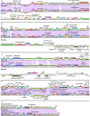
Genomic maps of B. cereus phages illustrate the high similarity of Shanette and JL contrasted to the unrelated phage Basilisk. Phages were mapped using Phamerator (19). Purple lines between phages denote regions of high nucleotide similarity, and the ruler shows genome base pairs. Boxes for gene products are labeled with predicted function, occasionally numbered, and colored to indicate similarity between the phages (E value < 1e−4). Predicted tRNAs are shown with black boxes and are labeled. DHFR, dihydrofolate reductase; MCP, major capsid protein; MurNAc-LAA, N-acetylmuramolyl-alanine amidase.
FIG 2
FIG 2
Whole-genome nucleotide (A) and amino acid (B) dot plot analysis of JL, Shanette. and Basilisk reveals relationships to other Bacillus phages and a clear division between JL/Shanette and Basilisk. Nucleotide dot plots were produced using Gepard (33) at a word length of 10 and amino acid at a word of 5. Whole-genome amino acid sequences were obtained from Phamerator (35).
FIG 3
FIG 3
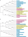
Phylogenetic trees of TerC (A) and TerD (B) produced by neighbor joining suggest that the JL and Shanette gene products are related to proteins encoded in Bacillus strains. All TerC and TerD proteins encoded by phages as well as a few representative Gram-positive and Gram-negative strains are shown (all have a BLASTP E value of greater than 10−7). Phylogenetic trees were constructed using a Muscle (28) alignment and the neighbor-joining method in Mega5 (29). Bootstrapping was set at 2000, and trees were collapsed at a less than 50% bootstrap value. str., strain.
FIG 4
FIG 4
Structural characterization of Basilisk, JL, and Shanette. (A) CsCl-purified phages run on SDS-polyacrylamide gels for virion component analysis. Basilisk (left lane) and JL (right lane) were run (the middle lane is empty). Lanes were cut into slices and analyzed by LC/MS/MS. Major proteins identified by high spectral count score and good coverage are indicated with their predicted molecular masses (in kilodaltons) at the sides of the gel. (B) Transmission electron microscopy confirms Basilisk as a Siphoviridae and JL and Shanette as Myoviridae. Plaque-purified phages were negatively stained and imaged.
FIG 5
FIG 5
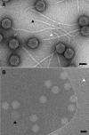
Transmission electron microscopy reveals two unique types of distal tail ends for Basilisk. (A) In a negative-stained preparation, tail striations were observed as well as two types of distal ends: a needle-like structure (black arrowhead) and a closed, rounded structure (white arrowhead). (B) In a cryogenic, unstained preparation, the tail striations are less visible because of the thin tails, unstained nature of the specimen, and low-electron-dose procedure used to record images. Partially empty (black arrowhead) and empty (white arrowhead) particles were also observed. Bars, 50 nm.
FIG 6
FIG 6
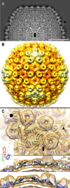
Three-dimensional reconstruction of the head of Basilisk phage from cryo-TEM images reveals an HK97-like fold. (A) A central slice perpendicular to an icosahedral 2-fold axis of symmetry (black oval). Protein and nucleic acid densities are represented with lighter intensities. The thin, corrugated outer ring is the capsid. Inside, a few layers of dsDNA are observed as concentric rings. Bar, 25 nm. (B) Surface rendering of the capsid viewed from the outside centered on an icosahedral 2-fold axis. (The contour selected corresponds to a sigma level of approximately 0.4.) Darker hues represent density closer to the center of the head. Icosahedral 5-fold axes lie in the center of the white five-pointed, star-shaped capsomeres (diameter of 78 nm). Between 5-fold and 2-fold (e.g., center of image) axes are hexamers with the appearance of a six- pointed star. A second type of hexameric capsomere is centered on the icosahedral 3-fold axes, which lie in the center of three adjacent 5-fold vertices. A lower-density feature was observed on 3-fold axes (see black arrowheads in panels A and B), which is likely not present at all 3-fold positions or is disordered. Between adjacent hexameric capsomeres and between hexameric and pentameric capsomeres are extensions from the capsid that have the appearance of donut-shaped trimers. (C) Fit of bacteriophage HK97 capsid protein core domain into cryo-EM reconstruction of the Basilisk capsid. The top view is from outside the capsid and is centered on one hexamer that is adjacent to the 5-fold symmetry axis. Threefold (triangle), 2-fold (oval), and 5-fold (pentagon) symmetry axes are marked. The bottom two views show slices through the capsid. The cryo-EM density is shown as an orange mesh. Black ribbons designate the nine subunits that form one asymmetric unit of the capsid. Blue ribbons show symmetrically related subunits. (Inset) HK97 capsid protein monomer (46) shown as a ribbon diagram. The N-terminal domain (yellow), cross-linking arm (green), A domain (blue), and P domain (red) are shown. As in a previous study (47), only the A and P domains were used in the fitting into the Basilisk capsid density. Coordinates omitted from the CW02 fitting (yellow, green, light red, and light blue) are indicated.

References

    1. Bergh O, Borsheim KY, Bratbak G, Heldal M. 1989. High abundance of viruses found in aquatic environments. Nature 340:467–468. 10.1038/340467a0 - DOI - PubMed
    1. Brussow H, Hendrix RW. 2002. Phage genomics: small is beautiful. Cell 108:13–16. 10.1016/S0092-8674(01)00637-7 - DOI - PubMed
    1. Hambly E, Suttle CA. 2005. The viriosphere, diversity, and genetic exchange within phage communities. Curr. Opin. Microbiol. 8:444–450. 10.1016/j.mib.2005.06.005 - DOI - PubMed
    1. Wilhelm SW, Jeffrey WH, Suttle CA, Mitchell DL. 2002. Estimation of biologically damaging UV levels in marine surface waters with DNA and viral dosimeters. Photochem. Photobiol. 76:268–273. 10.1562/0031-8655(2002)076<0268:EOBDUL>2.0.CO;2 - DOI - PubMed
    1. Wommack KE, Colwell RR. 2000. Virioplankton: viruses in aquatic ecosystems. Microbiol. Mol. Biol. Rev. 64:69–114. 10.1128/MMBR.64.1.69-114.2000 - DOI - PMC - PubMed

Publication types

LinkOut - more resources