. 2014 Nov;88(21):12242-53.
doi: 10.1128/JVI.01692-14. Epub 2014 Aug 6.

trans-Protease activity and structural insights into the active form of the alphavirus capsid protease

Affiliations

trans-Protease activity and structural insights into the active form of the alphavirus capsid protease

Megha Aggarwal et al. J Virol. 2014 Nov.

Abstract

The alphavirus capsid protein (CP) is a serine protease that possesses cis-proteolytic activity essential for its release from the nascent structural polyprotein. The released CP further participates in viral genome encapsidation and nucleocapsid core formation, followed by its attachment to glycoproteins and virus budding. Thus, protease activity of the alphavirus capsid is a potential antialphaviral target to arrest capsid release, maturation, and structural polyprotein processing. However, the discovery of capsid protease inhibitors has been hampered due to the lack of a suitable screening assay and of the crystal structure in its active form. Here, we report the development of a trans-proteolytic activity assay for Aura virus capsid protease (AVCP) based on fluorescence resonance energy transfer (FRET) for screening protease inhibitors. Kinetic parameters using fluorogenic peptide substrates were estimated, and the K(m) value was found to be 2.63 ± 0.62 μM while the k(cat)/K(m) value was 4.97 × 10(4) M(-1) min(-1). Also, the crystal structure of the trans-active form of AVCP has been determined to 1.81-Å resolution. Structural comparisons of the active form with the crystal structures of available substrate-bound mutant and inactive blocked forms of the capsid protease identify conformational changes in the active site, the oxyanion hole, and the substrate specificity pocket residues, which could be critical for rational drug design. IMPORTANCE The alphavirus capsid protease is an attractive antiviral therapeutic target. In this study, we have described the formerly unappreciated trans-proteolytic activity of the enzyme and for the first time have developed a FRET-based protease assay for screening capsid protease inhibitors. Our structural studies unveil the structural features of the trans-active protease, which has been previously proposed to exist in the natively unfolded form (M. Morillas, H. Eberl, F. H. Allain, R. Glockshuber, and E. Kuennemann, J. Mol. Biol. 376:721-735, 2008, doi:http://dx.doi.org/10.1016/j.jmb.2007.11.055). The different enzymatic forms have been structurally compared to reveal conformational variations in the active and substrate binding sites. The flexible active-site residue Ser218, the disordered C-terminal residues after His261, and the presence of a water molecule in the oxyanion hole of AVCPΔ2 (AVCP with a deletion of the last two residues at the C terminus) reveal the effect of the C-terminal Trp267 deletion on enzyme structure. New structural data reported in this study along with the fluorogenic assay will be useful in substrate specificity characterization, high-throughput protease inhibitor screening, and structure-based development of antiviral drugs.


Figures

FIG 1
FIG 1
Purification and trans-proteolytic activity analysis for AVCPΔ2. (A) Schematic representation of inactive and active forms of alphavirus CP. In the inactive form Trp267 shows auto-inhibitory action on enzyme activity after autoproteolysis (left). In the absence of Trp267, the protein acquires its catalytic property as the substrate can easily access the active site (right). (B) Gel filtration chromatography and SDS-PAGE analysis of major peak fractions show the protein (AVCPΔ2) purified to homogeneity. The protein is monomeric in nature, as determined by the gel filtration profile. AU, arbitrary units. (C) Analysis of in vitro trans-proteolytic activity of AVCPΔ2. The enzymatic cleavage assay was carried out using 15 μg of protein in HEPES buffer (20 mM, pH 7.0) by the addition of 1 μM fluorogenic peptide substrate at room temperature. The hydrolysis of the peptide substrate was measured at different time intervals. The excitation was done at 340 nm, and the emission spectrum for each time point is shown as a scan from 450 nm to 600 nm. (D) The initial velocity (μM/min) was calculated for increasing concentrations of the substrate. Different substrate (S) concentrations ranging from 0.6 μM to 16 μM were used (x axis). The experiment was done in triplicate, and the values represent the average data. All the data were normalized using the same reaction mixture without the enzyme.
FIG 2
FIG 2
Crystal structure of AVCPΔ2 and its comparison with other enzymatic forms of the alphavirus CP. (A) Overall structure of the monomer contains two β-barrel subdomains consisting of the catalytic triad in between the cleft. The catalytic triad residues are presented in sticks. (B) Surface view of AVCPΔ2 showing different pockets and regions involved in catalysis, represented with different colors. (C) Both chains of the active (chain A in green and chain B in yellow color) and substrate-bound (chain A in blue and chain B in gray) forms were aligned along with native AVCP (pink). Circle 1 (black) shows the variation in the C-terminal region in two chains of the substrate-bound form, while this region is absent from the active AVCP and remains intact in native AVCP. Circle 2 (blue) shows the loop flexibility in the S1 specificity pocket. (D) Closeup view of the dimeric interaction in AVCPΔ2. The residues involved in crystallographic dimer formation are shown as sticks. Asn225 of one chain forms H-bonds with Phe191 and Tyr192 of the other chain. Chain B residues are labeled with a prime (′) sign. Chain A is shown in green while chain B is in yellow.
FIG 3
FIG 3
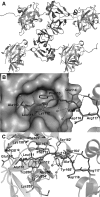
The hydrophobic pocket of AVCPΔ2 with the bound N-terminal arm of the neighboring molecule. (A) The crystal structure of AVCPΔ2 dimer with the symmetry-related molecules. The hydrophobic pocket of each molecule occupied with the N-terminal arm of the neighboring symmetry-related molecule is shown. The dotted square highlights one such binding. (B) The zoomed view of the hydrophobic pocket of one of the AVCPΔ2 subunits with the bound N-terminal arm of the neighboring subunit is displayed in the surface view. (C) Cartoon view. All of the polar interactions are shown with dotted lines, and the interacting water molecules are shown as spheres.
FIG 4
FIG 4
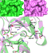
Structural comparison of the hydrophobic pockets between native and active forms of AVCP. The surface view (A) and the superposed view of the hydrophobic pocket of the active AVCPΔ2 (green) over the inactive AVCP (pink) crystal structure (B), which shows the differences in the side-chain conformation of some of the residues that occur at this hydrophobic pocket to accommodate the N-terminal arm of the neighboring subunit.
FIG 5
FIG 5
Comparative studies of native and active AVCP for structural differences in the catalytic triad and S1 specificity pocket. (A) The catalytic triad residues from different enzymatic states: native AVCP (pink), active AVCPΔ2 chain A (green), and AVCPΔ2 chain B (yellow) show conformational changes in Ser218 side chain. The catalytic triad residues and Trp267 are shown as sticks. The interactions of Ser218 and His144 with Trp267 are shown as red dashed lines. (B) The differences at the S1 specificity pocket of AVCPΔ2 (green) are shown in superposition with the native AVCP (pink) crystal structure with bound Trp267 at the active site. The dotted red lines highlight the difference at region Gly213 to Gly216. The red dotted box highlights the flipped peptide bond at Pro215 and Gly216. (C) The differences in the volume of the S1 specificity pocket encircled by a dotted red circle are shown in surface view.
FIG 6
FIG 6
Oxyanion hole of different enzymatic forms of alphavirus CP. In panels A and B, the oxyanion hole of AVCPΔ2 and chain A of substrate-bound SCP is occupied by a water molecule. Panels C and D, in contrast, represent the H bonding of oxyanion hole residues with the carbonyl oxygen atom preceding the scissile bond in chain B of the substrate-bound form and native AVCP, respectively.
FIG 7
FIG 7
Structural comparison of substrate specificity pockets of native and active forms of AVCP. (A) Specificity pocket S1 of AVCPΔ2 chain A (green) and chain B (yellow) is compared with that of native AVCP (pink). The main chain backbone at the region of Gly213 to Gly214 shows major differences in both chain A (2.9 Å) and chain B (3.4 Å) compared to the native form. Gly236 interacts with Val265, Asn238 interacts with Thr210, and Leu245 interacts with His261 in the native form, and these interactions are absent in AVCPΔ2. (B) Leu234 main chain forms an H bond with Ser218 as well as Trp267. (C) Comparison of the S4 pocket shows deviation in the backbone as well as side chains. Ser246 interaction with His261 and Thr264 is found in inactive AVCP but is absent from the active form. (D) Comparison of the S4′ and S2′ pockets in active and inactive forms of AVCP demonstrates the displacement of 0.5, 0.9, and 0.6 Å in Asn123, Lys127, and Ile128, respectively.

References

    1. Strauss JH, Strauss EG. 1994. The alphaviruses: gene expression, replication, and evolution. Microbiol. Rev. 58:491–562. - PMC - PubMed
    1. Choi HK, Tong L, Minor W, Dumas P, Boege U, Rossmann MG, Wengler G. 1991. Structure of Sindbis virus core protein reveals a chymotrypsin-like serine proteinase and the organization of the virion. Nature 354:37–43. 10.1038/354037a0. - DOI - PubMed
    1. Melancon P, Garoff H. 1987. Processing of the Semliki Forest virus structural polyprotein: role of the capsid protease. J. Virol. 61:1301–1309. - PMC - PubMed
    1. Weiss B, Nitschko H, Ghattas I, Wright R, Schlesinger S. 1989. Evidence for specificity in the encapsidation of Sindbis virus RNAs. J. Virol. 63:5310–5318. - PMC - PubMed
    1. Hahn CS, Lustig S, Strauss EG, Strauss JH. 1988. Western equine encephalitis virus is a recombinant virus. Proc. Natl. Acad. Sci. U. S. A. 85:5997–6001. 10.1073/pnas.85.16.5997. - DOI - PMC - PubMed

Publication types

MeSH terms

Associated data

LinkOut - more resources