Review
. 2014:2014:154631.
doi: 10.1155/2014/154631. Epub 2014 Jul 2.

Thalamic lesions: a radiological review

Affiliations
Review

Thalamic lesions: a radiological review

Dimitri Renard et al. Behav Neurol. 2014.

Abstract

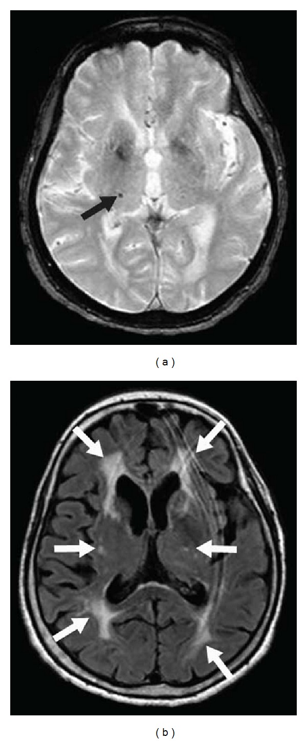
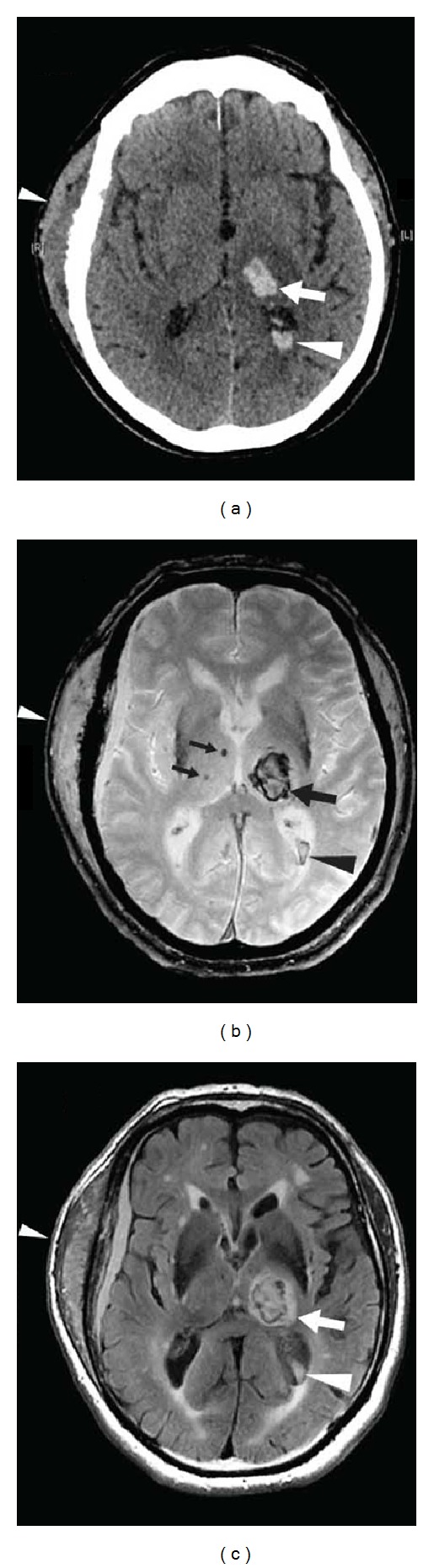
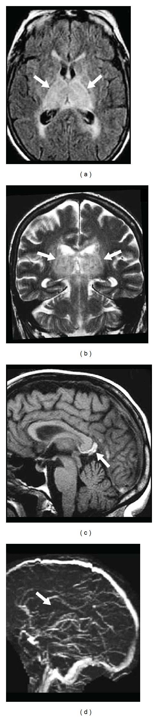
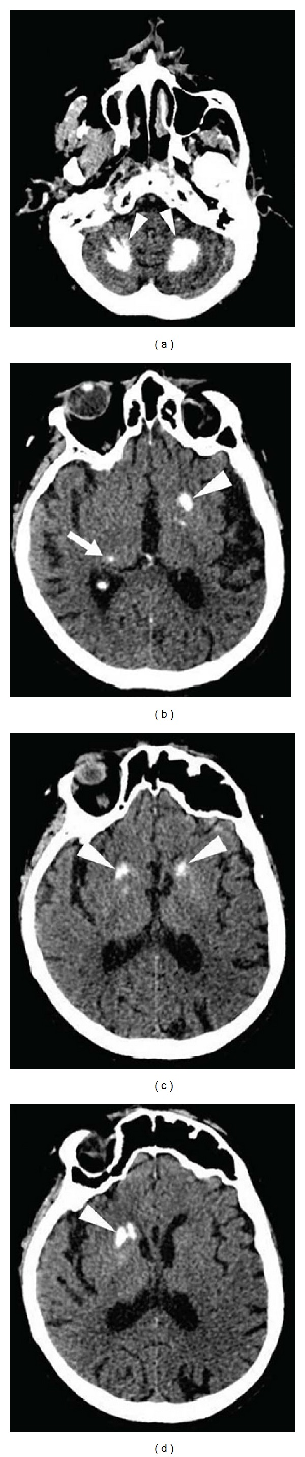
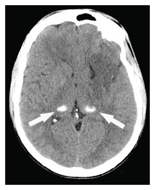
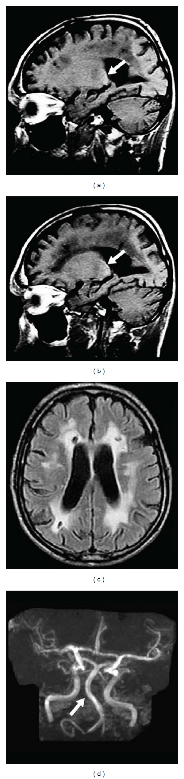
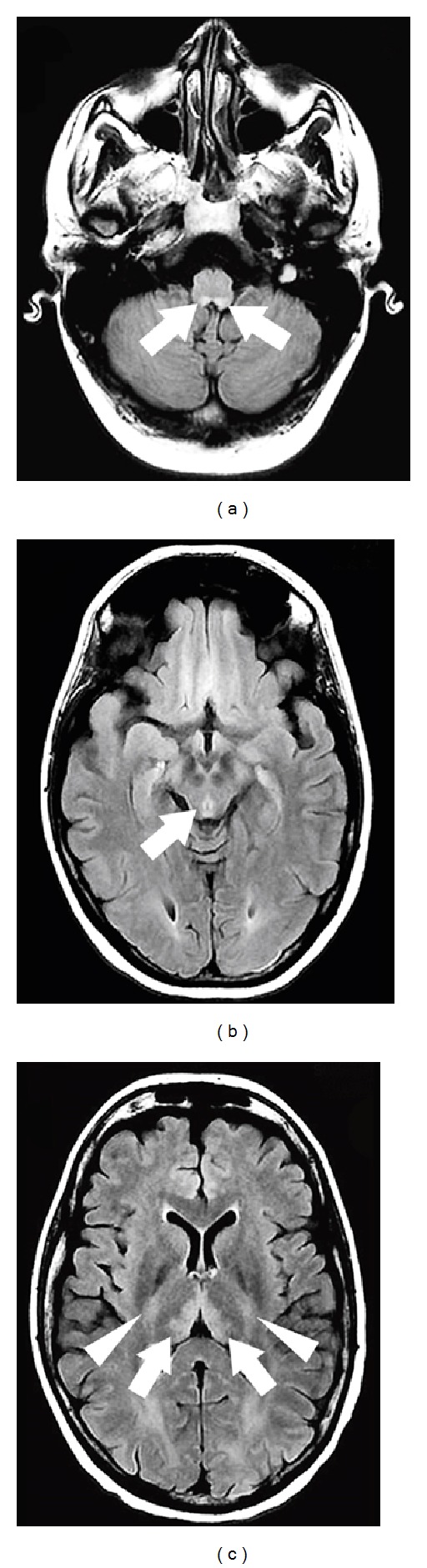
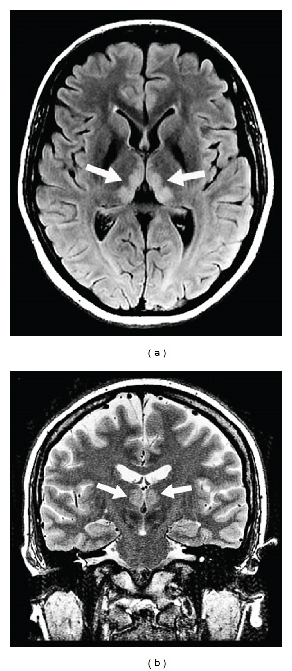
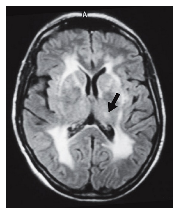
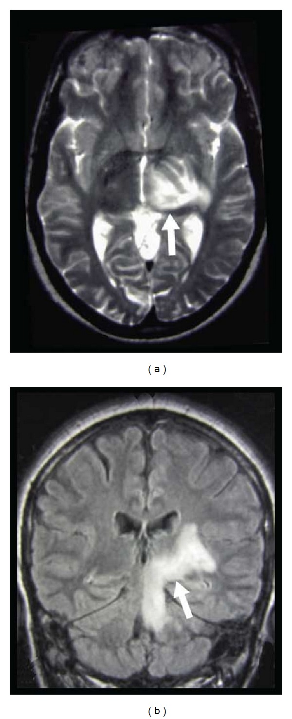
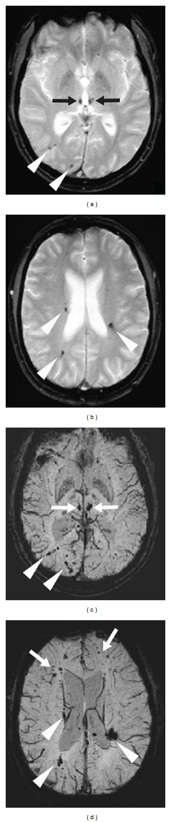
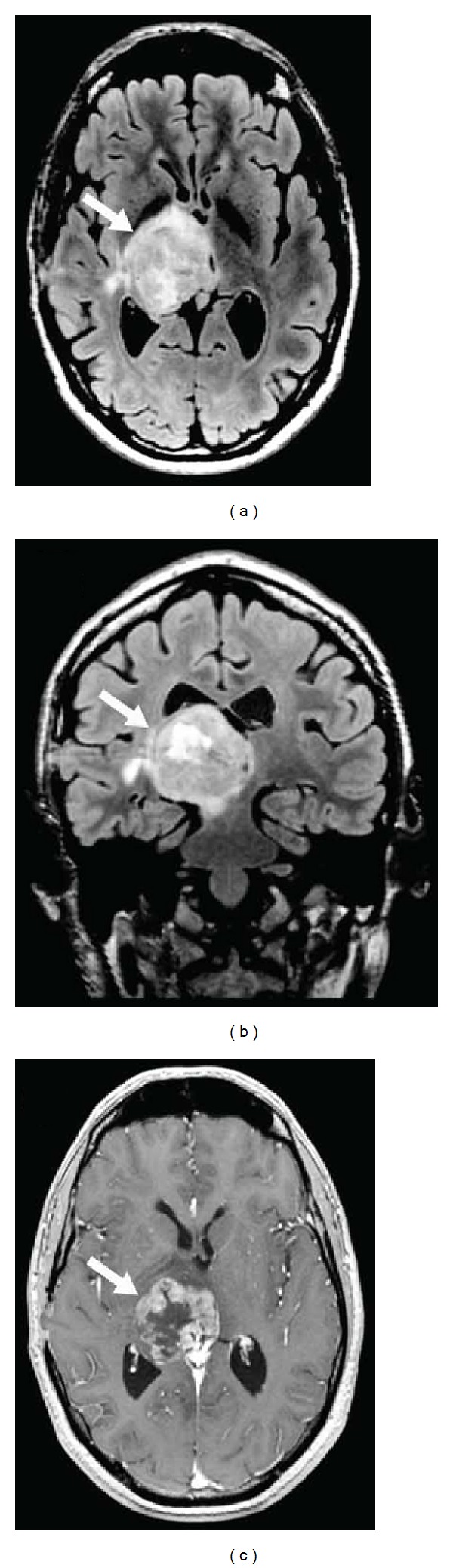
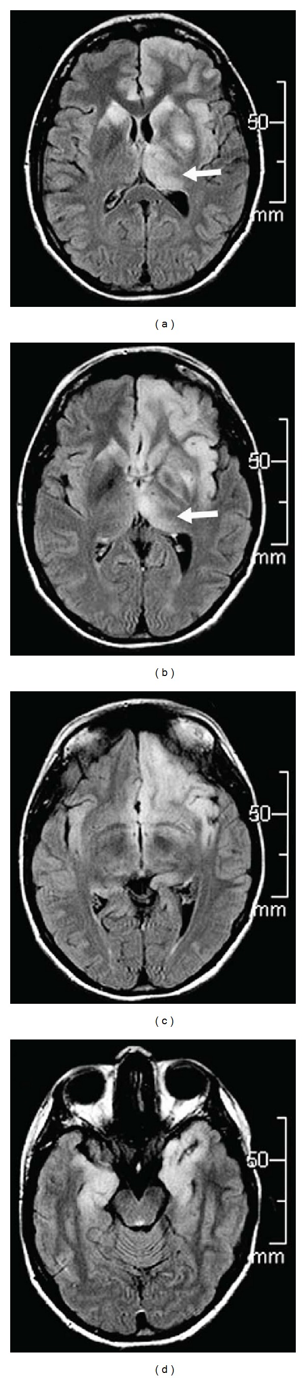
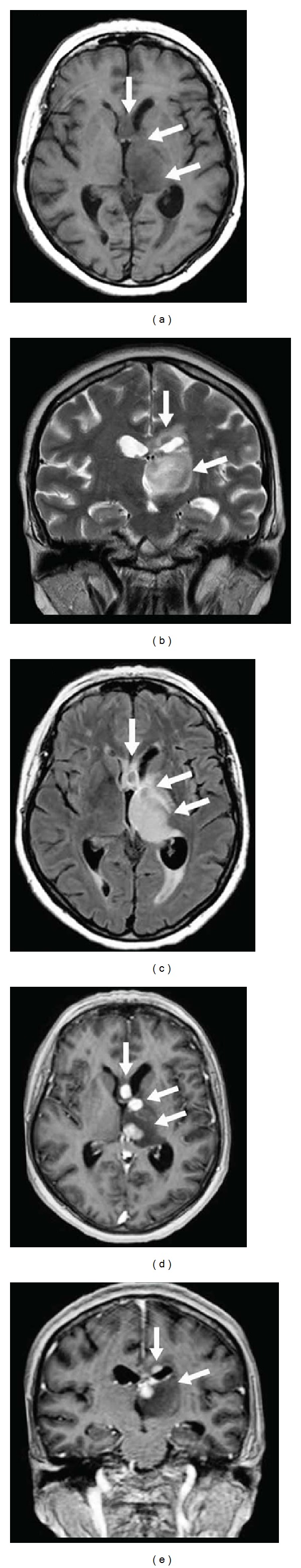
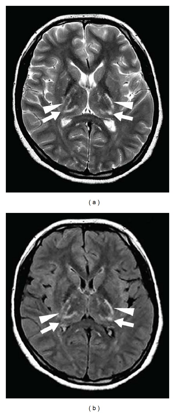
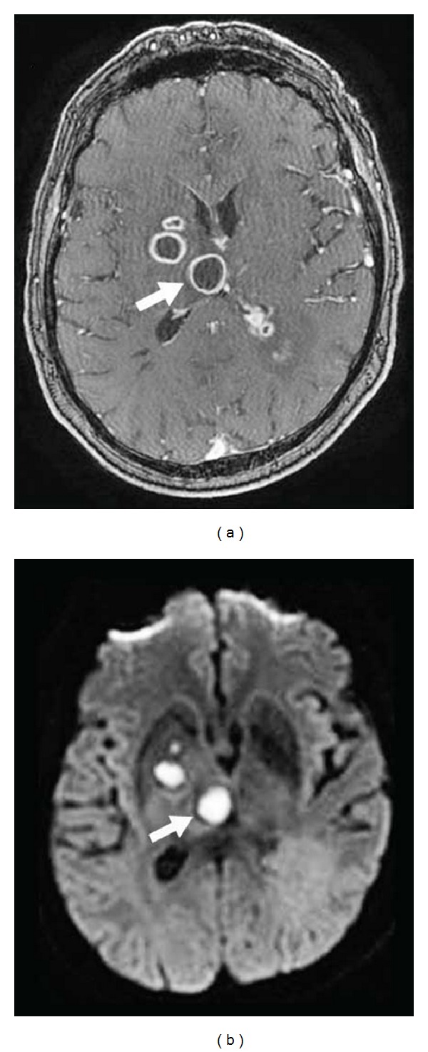
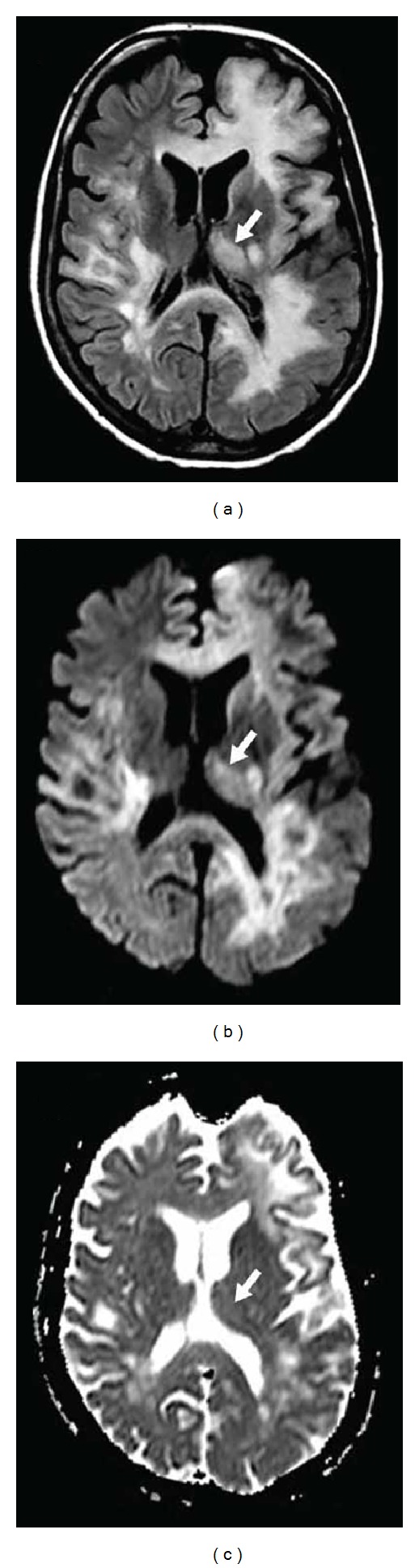
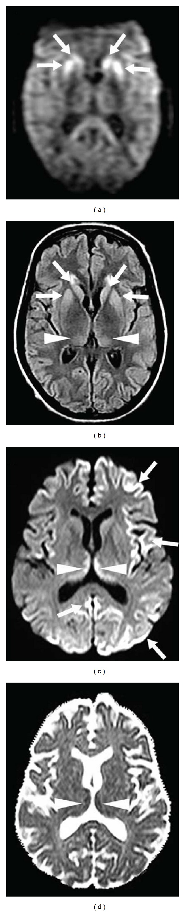
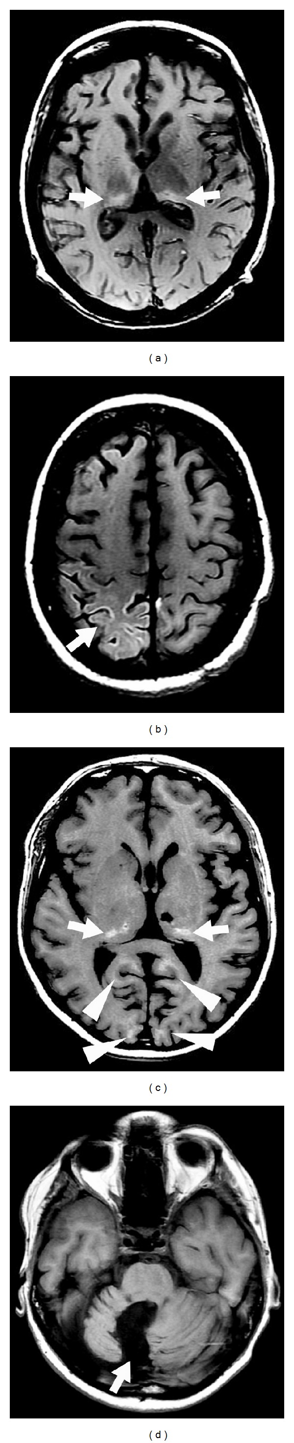
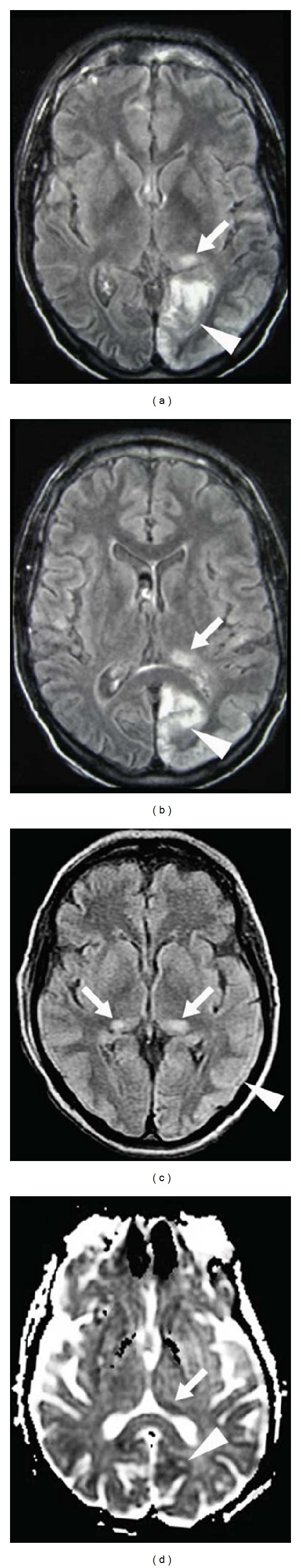
Background: Thalamic lesions are seen in a multitude of disorders including vascular diseases, metabolic disorders, inflammatory diseases, trauma, tumours, and infections. In some diseases, thalamic involvement is typical and sometimes isolated, while in other diseases thalamic lesions are observed only occasionally (often in the presence of other typical extrathalamic lesions).

Summary: In this review, we will mainly discuss the MRI characteristics of thalamic lesions. Identification of the origin of the thalamic lesion depends on the exact localisation inside the thalamus, the presence of extrathalamic lesions, the signal changes on different MRI sequences, the evolution of the radiological abnormalities over time, the history and clinical state of the patient, and other radiological and nonradiological examinations.


Figures

Figure 1
Figure 1
Schematic representation of the blood supply to the thalamus.
Figure 2
Figure 2
Thalamic infarctions in the thalamogeniculate artery territory (arrows in a, d, and g) in 3 different patients (patient 1 in a, b, and c; patient 2 in d, e, and f; patient 3 in g, h, and i) associated with other infarction areas in the ipsilateral posterior cerebral artery territories (arrowheads on a, b, d, e, g, and h) seen on DWI imaging due to posterior cerebral artery occlusion seen on TOF sequences (arrows on c, f, and i).
Figure 3
Figure 3
DWI (a) and FLAIR (b and c) imaging showing infarctions involving the territories of the right-sided posterior choroidal artery (a and b, arrows), the posterior cerebral artery (a, b, and c, arrowheads), the collicular artery, and the posteromedial choroidal artery (c, arrows).
Figure 4
Figure 4
DWI (a and c) and ADC map (b and d) of 2 different patients (patient 1, a and b; patient 2, c and d) showing paramedian anterior thalamic infarction, bilateral in patient 1 and unilateral in patient 2.
Figure 5
Figure 5
Gradient-echo (a) and FLAIR (b) sequences showing a right-sided thalamic microbleed (a) associated with diffuse ischemic white matter hyperintensities on FLAIR imaging (b) in a patient with severe and chronic arterial hypertension.
Figure 6
Figure 6
CT scan (a) and gradient-echo-weighted (b) and FLAIR (c) imaging showing a hypertension-related left-sided thalamic hemorrhage (large arrows on a, b, and c) and surrounding edema, complicated with a intraventricular hemorrhage (large arrowheads on a, b, and c) and associated with chronic hypertension-related microbleeds in the right thalamus (small arrows on b) and a subcutaneous and subdural hemorrhage (small arrowheads on a, b, and c) related to trauma (caused by acute right hemiplegie due to the thalamic hemorrhage).
Figure 7
Figure 7
Bilateral thalamic vasogenic edema seen as hyperintensity on both axial FLAIR (a) and coronal T2-weighted (b) imaging due to venous thrombosis of the deep cerebral venous system. The venous thrombosis of the vein of Galen is seen as hyperintensity on sagittal unenhanced T1-weighted imaging (c), and lack of flow in the deep cerebral venous system is seen on MR venography (d).
Figure 8
Figure 8
A patient with vitamin D deficiency with extensive cerebellar (a) and basal ganglia (arrowheads on b, c, and d) and thalamic (arrow on b) calcifications on CT.
Figure 9
Figure 9
A patient with atypical Werner syndrome (i.e., a progeroid syndrome with Werner syndrome phenotype but without typical RECQL2 mutation) due to a LMNA mutation showing bilateral thalamic calcifications on CT scan.
Figure 10
Figure 10
Sagittal unenhanced T1-weighted imaging (a and b) in a Fabry patient showing hyperintensity in the pulvinar, associated with ischemic leukoencephalopathy on FLAIR sequences (c) and vertebrobasilar dolichoectasia on TOF imaging (d).
Figure 11
Figure 11
A patient with Wernicke syndrome showing FLAIR hyperintensities in the hypoglossal nuclei (a), the periaqueductal gray matter and mesencephalic tectum (b), the medial part of the thalami (arrows, c), and the pyramidal tracts (arrowheads, c).
Figure 12
Figure 12
A patient with Wernicke syndrome showing hyperintensities in the medial part of the thalami on axial FLAIR (a) and coronal T2-weighted (b) imaging.
Figure 13
Figure 13
A patient with preeclampsia-related reversible posterior leukoencephalopathy syndrome with cortical and subcortical (arrowhead on a, b, c, and d) vasogenic edema seen as hyperintensity on FLAIR sequences also present in the left thalamus (arrow, a).
Figure 14
Figure 14
A patient with longstanding multiple sclerosis with diffuse leukoencephalopathy and a left thalamic demyelinating lesion (a) on FLAIR imaging.
Figure 15
Figure 15
A patient with neuro-Behcet showing a left-sided hyperintense lesion on axial T2-weighted (a) and coronal FLAIR (b) imaging involving the cerebellum, the midbrain, and the inferior part of the thalamus (arrows), the internal capsule, and the putamen.
Figure 16
Figure 16
A patient after a major head trauma showing diffuse axonal injury with small hemorrhagic lesions in the medial part of both thalami (arrows, a) and multiple lesions near the cortico-subcortical junction (arrowheads, a and b) seen as hypointensity on gradient-echo-weighted imaging (a and b). Multiple additional lesions can be seen on SWI imaging (c and d) demonstrating the superiority of SWI imaging in diffuse axonal injury.
Figure 17
Figure 17
A patient with glioblastoma multiforme in the right thalamus seen as hyperintensity on axial (a) and coronal (b) FLAIR imaging, with gadolinium-enhancement on axial T1-weighted imaging (c).
Figure 18
Figure 18
A patient with gliomatosis cerebri involving the left thalamus (arrows, a and b), the bilateral basal ganglia (a and b), the left frontal lobe (a, b, and c), and both temporal lobes (d), seen as hyperintensity on FLAIR sequences.
Figure 19
Figure 19
A patient with primary CNS lymphoma involving the left-sided thalamus, the internal capsule, and the anterior part of the corpus callosum seen as hypointensity on axial unenhanced T1-weighted imaging (a), hyperintensity on coronal T2-weighted (b) and axial FLAIR (c) imaging, with multifocal enhancement on axial (d) and coronal (e) gadolinium-enhanced T1-weighted imaging.
Figure 20
Figure 20
A patient with Mycoplasma pneumoniae-related acute necrotizing encephalitis involving symmetrically the posterior part of the internal capsule (arrowheads) and the posterolateral portion of the thalamus (arrows) on both sides, seen as hyperintensity on both T2-weighted (a) and FLAIR (b) imaging.
Figure 21
Figure 21
A patient with several brain abscesses involving the right thalamus (arrows) and the basal ganglia, with ring enhancement on gadolinium-enhanced T1-weighted imaging (a) and restricted diffusion seen as hyperintensity on DWI imaging (b).
Figure 22
Figure 22
Diffuse white matter and associated left frontal cortical and left thalamic (arrows) involvement seen as hyperintensity on FLAIR (a), DWI (b), and ADC map (c) in a patient with natalizumab-related (for multiple sclerosis) progressive multifocal leukoencephalopathy.
Figure 23
Figure 23
Two different sporadic CJD patients (patient 1, a and b; patient 2, c and d) showing in patient 1 bilateral caudate nucleus and putaminal (arrows) and to a lesser degree posteromesial and pulvinar thalamic (arrowheads) hyperintensities on DWI (a) and FLAIR imaging (b) and in patient 2 bilateral posteromesial thalamic (arrowheads) and multifocal cortical (arrows) hyperintensities on DWI (c), seen as hypointensity on ADC map (d). Anterior predominance of the thalamic involvement can be seen in patient 2.
Figure 24
Figure 24
T1-weighted MRI of patient 1 (a and b) after generalized status epilepticus and patient 2 (c and d) with a history of cerebellar astrocytoma treated by surgical resection and large field radiation therapy including the occipital lobes and both thalami showing laminar necrosis of both thalami (a and c, arrows) associated with laminar necrosis in the right-sided parietal cortex (b, arrows) and in the bilateral occipital cortex (c, arrowheads). Panel (d) shows the cerebellar cavity due to cerebellar astrocytoma resection.
Figure 25
Figure 25
Peri-ictal thalamic lesions (arrows) following status epilepticus in patient 1 (a and b) with partial occipital status epilepticus related to MELAS in the left occipital lobe (arrowheads on a and b) all seen as hyperintensity on FLAIR imaging and in patient 2 (c and d) with generalized status epilepticus with also left occipitotemporal cortical signal changes due to the seizures seen as hyperintensity on FLAIR imaging (c) and as hypointensity on ADC map (d).

Similar articles

Cited by

References

    1. Schmahmann JD. Vascular syndromes of the thalamus. Stroke. 2003;34:2264–2278. - PubMed
    1. Vinters HV. Cerebral amyloid angiopathy. A critical review. Stroke. 1987;18:311–324. - PubMed
    1. de Champfleur NM, Langlois C, Ankenbrandt WJ, et al. Magnetic resonance imaging evaluation of cerebral cavernous malformations with susceptibility-weighted imaging. Neurosurgery. 2011;68:641–647. - PubMed
    1. Moore DF, Ye F, Schiffmann R, Butman JA. Increased signal intensity in the pulvinar on T1-weighted images: a pathognomonic MR imaging sign of Fabry disease. American Journal of Neuroradiology. 2003;24:1096–1101. - PMC - PubMed
    1. Reisin RC, Romero C, Marchesoni C, et al. Brain MRI findings in patients with Fabry disease. Journal of the Neurological Sciences. 2011;305(1):41–44. - PubMed

MeSH terms

LinkOut - more resources