

Thalamic lesions: a radiological review
- PMID: 25100900
- PMCID: PMC4101959
- DOI: 10.1155/2014/154631
Thalamic lesions: a radiological review
Abstract
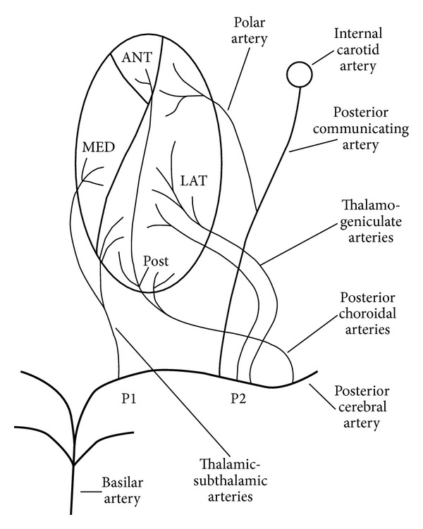
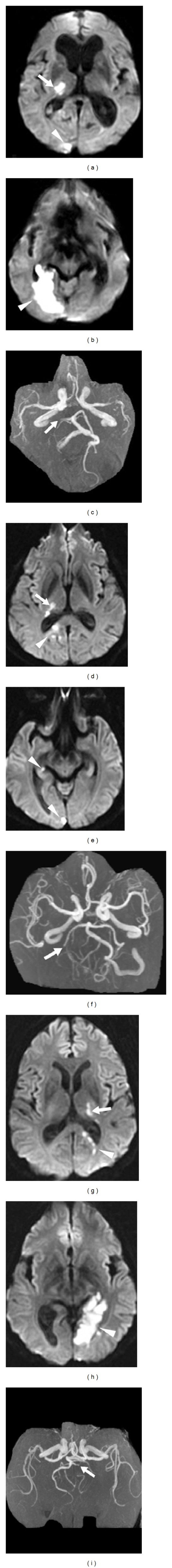
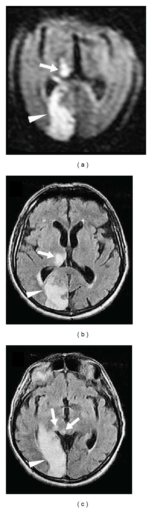
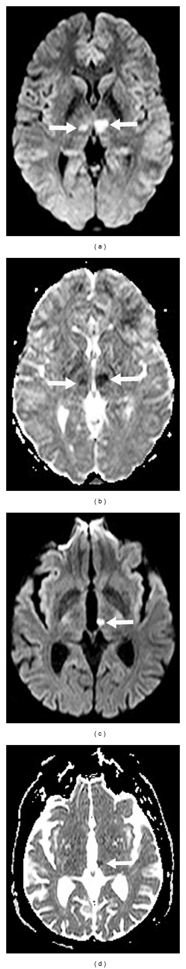
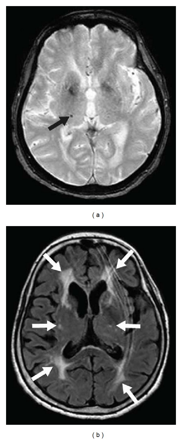
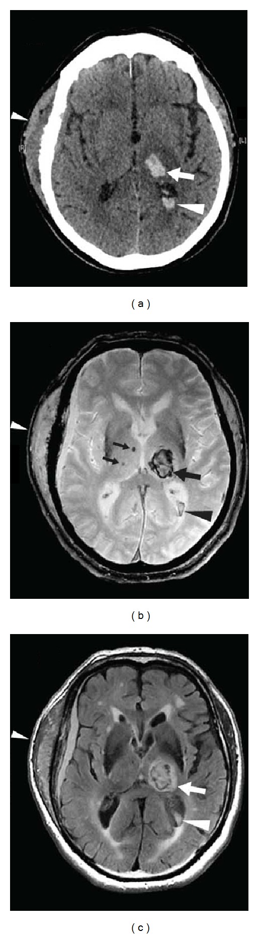
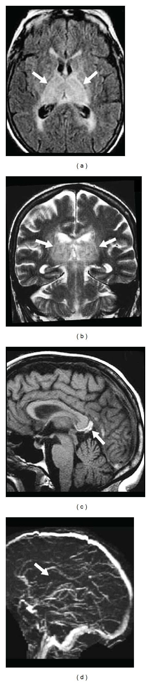
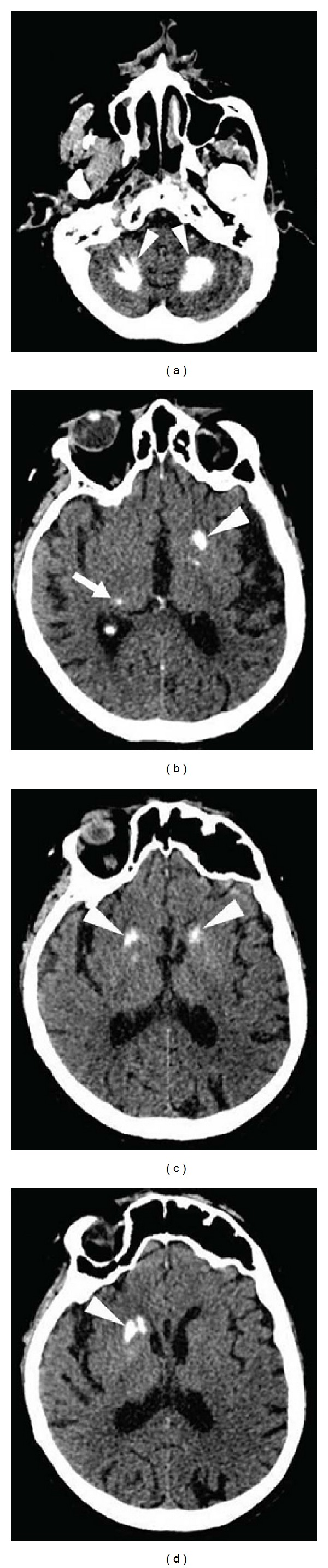
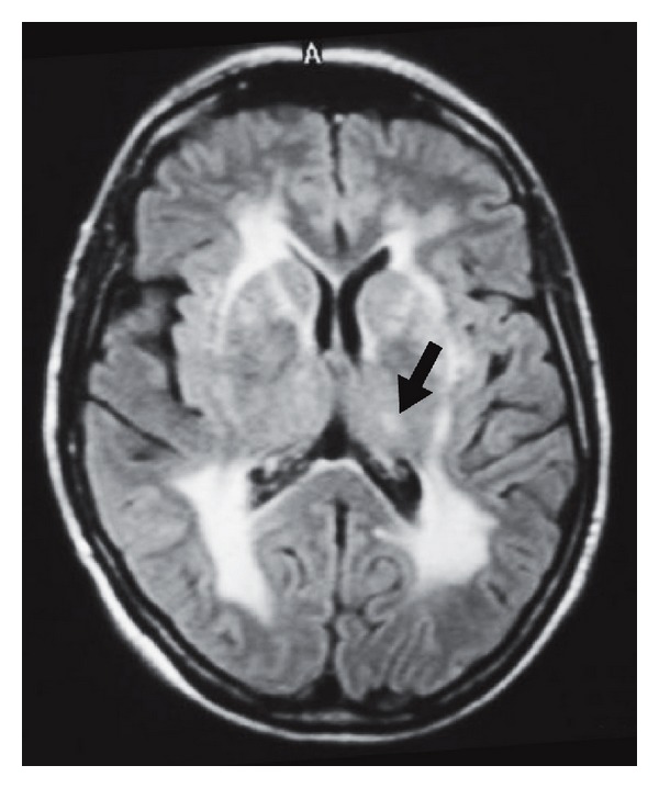
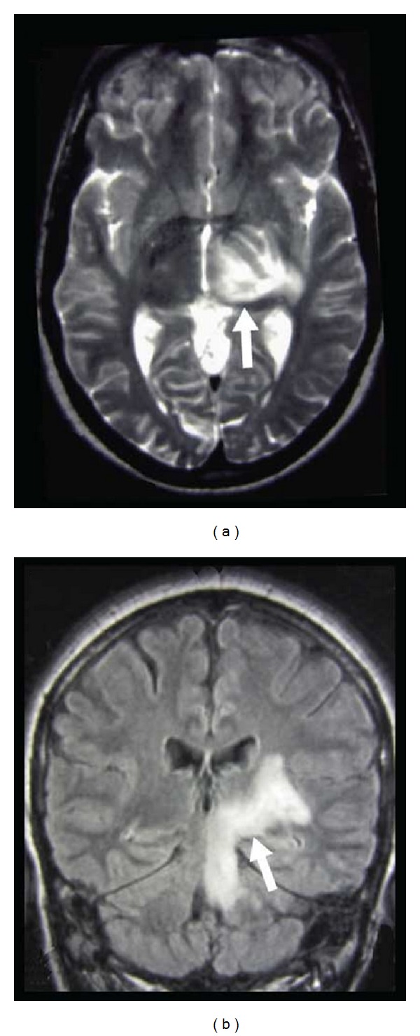
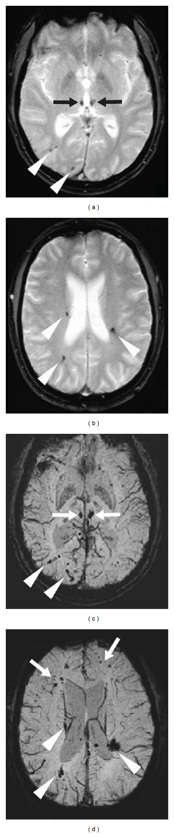
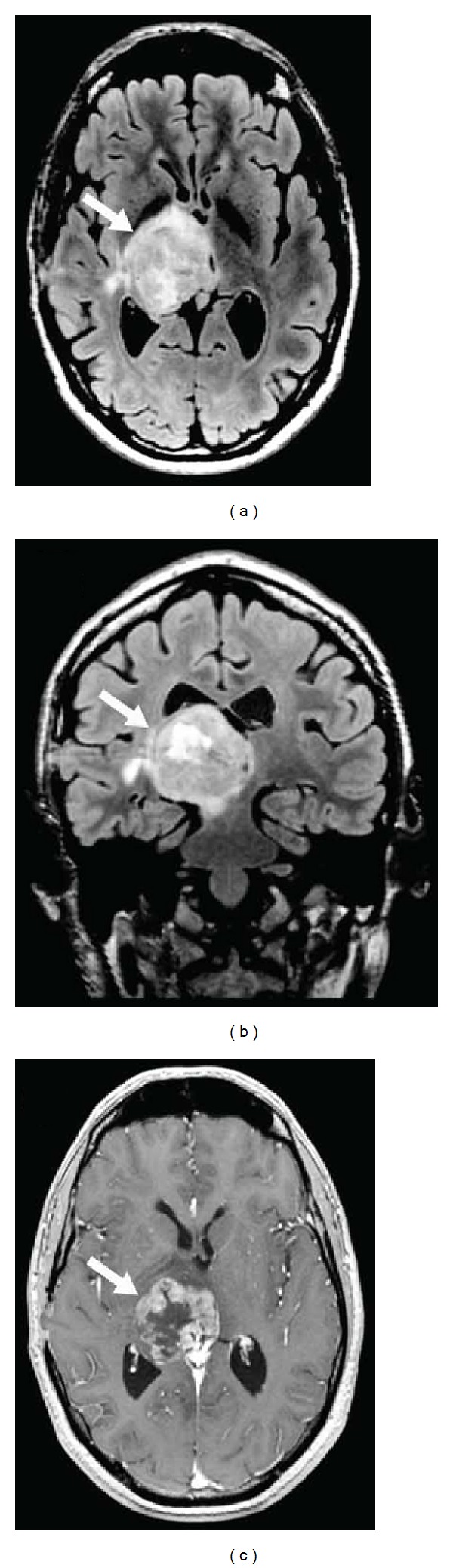
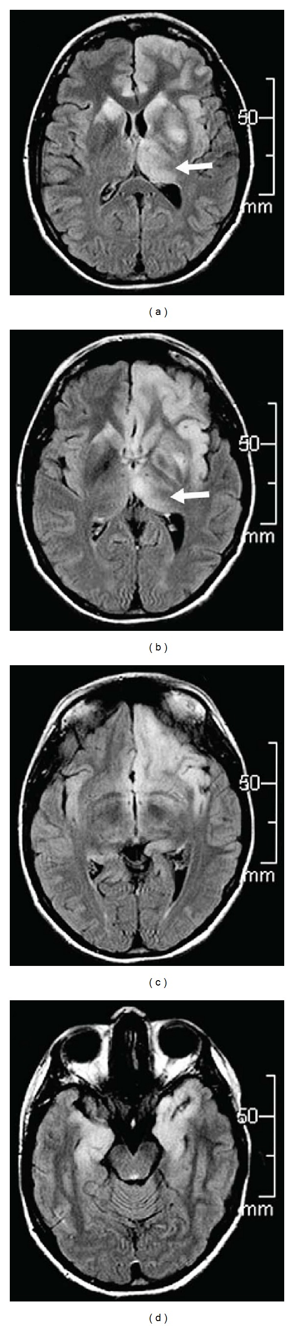
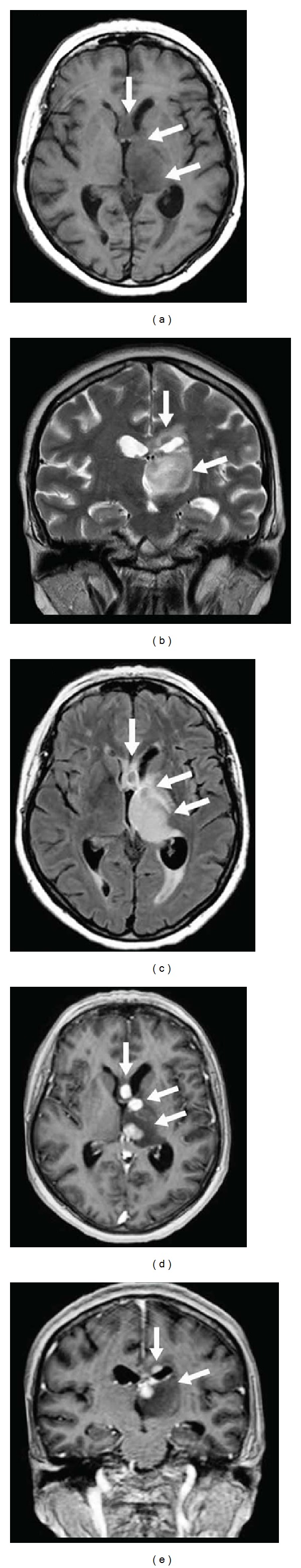
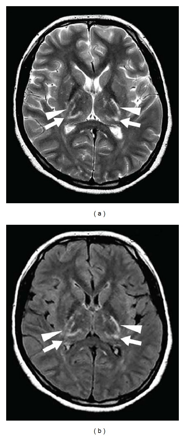
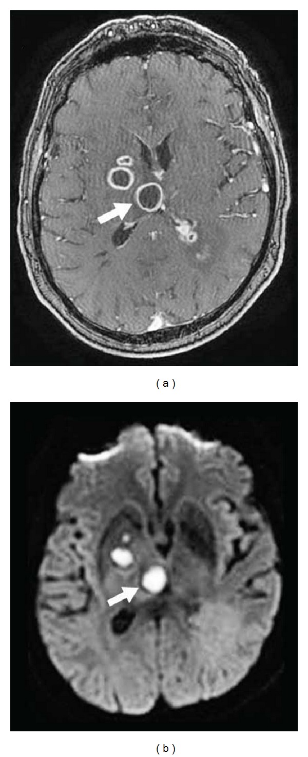
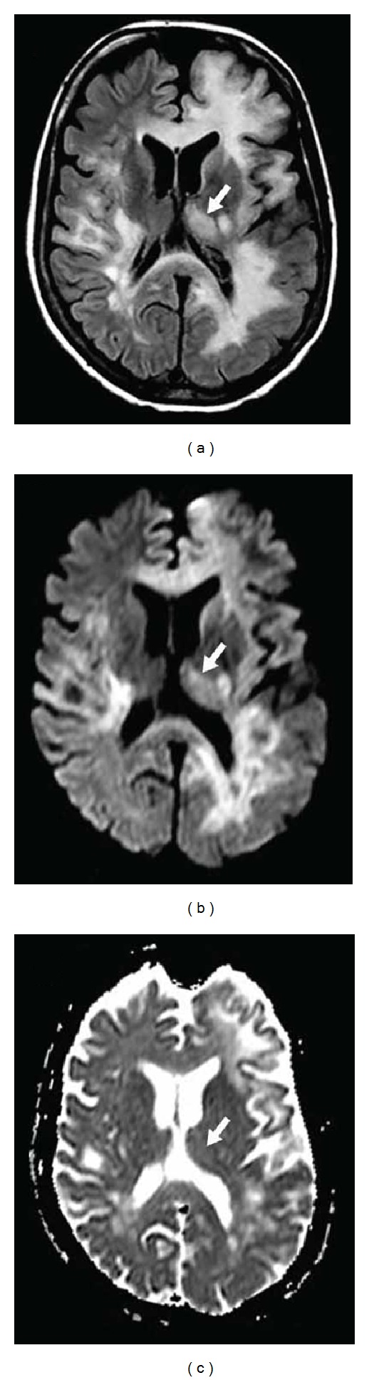
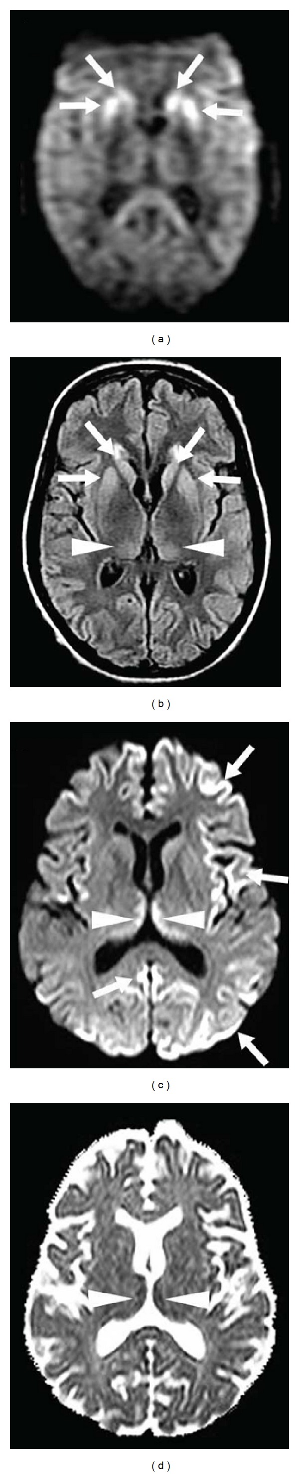
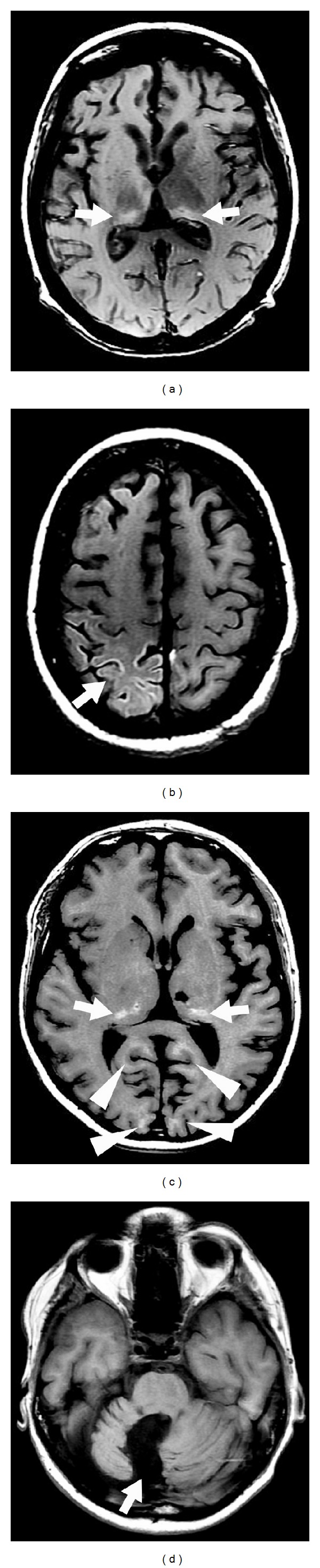
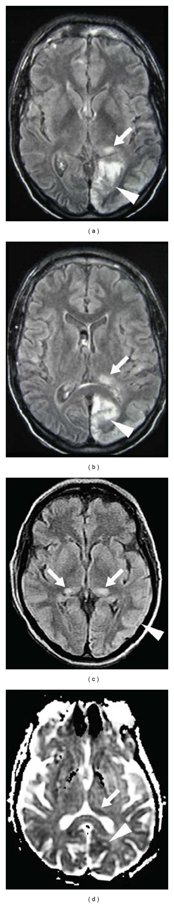
Background: Thalamic lesions are seen in a multitude of disorders including vascular diseases, metabolic disorders, inflammatory diseases, trauma, tumours, and infections. In some diseases, thalamic involvement is typical and sometimes isolated, while in other diseases thalamic lesions are observed only occasionally (often in the presence of other typical extrathalamic lesions).
Summary: In this review, we will mainly discuss the MRI characteristics of thalamic lesions. Identification of the origin of the thalamic lesion depends on the exact localisation inside the thalamus, the presence of extrathalamic lesions, the signal changes on different MRI sequences, the evolution of the radiological abnormalities over time, the history and clinical state of the patient, and other radiological and nonradiological examinations.
Figures

References
-
- Schmahmann JD. Vascular syndromes of the thalamus. Stroke. 2003;34:2264–2278. - PubMed
-
- Vinters HV. Cerebral amyloid angiopathy. A critical review. Stroke. 1987;18:311–324. - PubMed
-
- de Champfleur NM, Langlois C, Ankenbrandt WJ, et al. Magnetic resonance imaging evaluation of cerebral cavernous malformations with susceptibility-weighted imaging. Neurosurgery. 2011;68:641–647. - PubMed
-
- Reisin RC, Romero C, Marchesoni C, et al. Brain MRI findings in patients with Fabry disease. Journal of the Neurological Sciences. 2011;305(1):41–44. - PubMed
Publication types
MeSH terms
LinkOut - more resources
Full Text Sources
Other Literature Sources
