

Review of choroidal osteomas
- PMID: 25100910
- PMCID: PMC4123278
- DOI: 10.4103/0974-9233.134686
Review of choroidal osteomas
Abstract
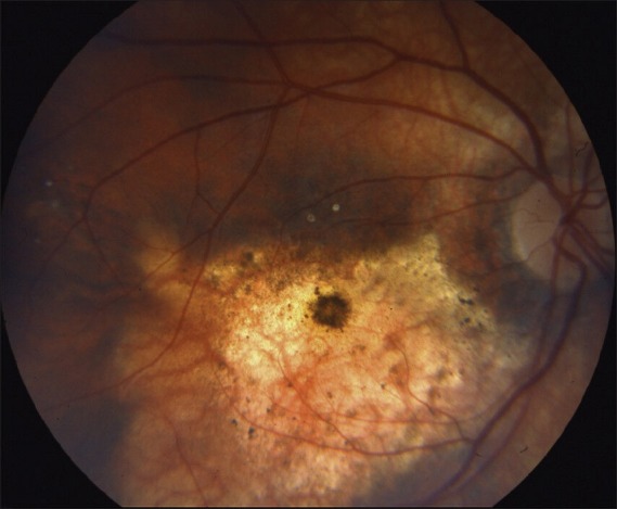
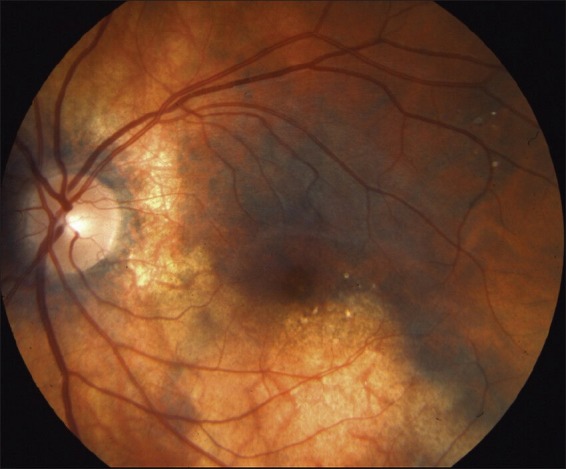
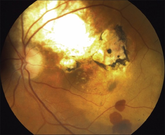
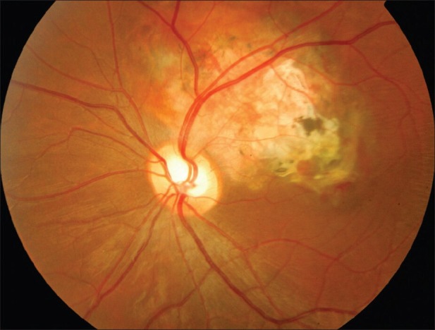
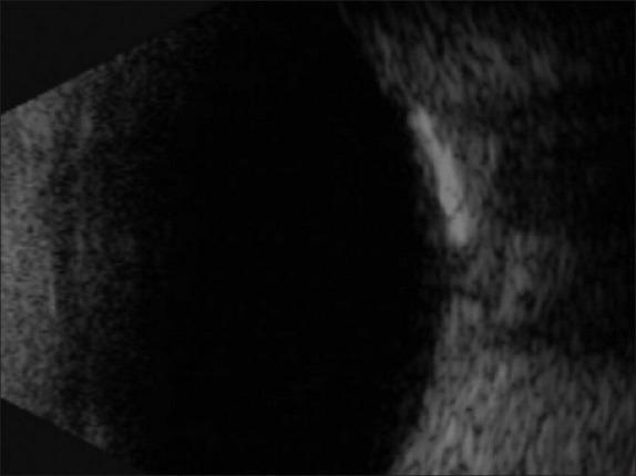
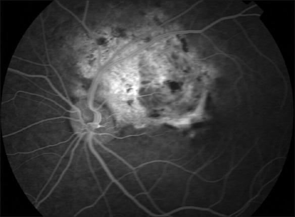
Choroidal osteomas are rare benign ossifying tumors that appear as irregular slightly elevated, yellow-white, juxtapapillary, choroidal mass with well-defined geographic borders, depigmentation of the overlying pigment epithelium; and with multiple small vascular networks on the tumor surface. Visual loss results from three mechanisms: Atrophy of the retinal pigment epithelium overlying a decalcified osteoma; serous retinal detachment over the osteoma from decompensated retinal pigment epithelium, and most commonly from choroidal neovascularization. Recent evidence points to the beneficial effects of intravitreal vascular endothelial growth factor antagonists in improving visual acuity in serous retinal detachment with or without choroidal neovascularization.
Keywords: Argon Laser; Choroidal Osteoma; Intravitreal Bevacizumab; Intravitreal Ranibizumab; Photodynamic Therapy.
Conflict of interest statement
Figures
References
-
- Gass JD, Guerry RK, Jack RL, Harris G. Choroidal osteoma. Arch Ophthalmol. 1978;96:428–35. - PubMed
-
- Williams AT, Font RL, Van Dyk HJ, Riekhof FT. Osseous choristoma of the choroid simulating a choroidal melanoma. Association with a positive 32 P test. Arch Ophthalmol. 1978;96:1874–7. - PubMed
-
- Shields CL, Sun H, Demirci H, Shields JA. Factors predictive of tumor growth, tumor decalcification, choroidal neovascularization, and visual outcome in 74 eyes with choroidal osteoma. Arch Ophthalmol. 2005;123:1658–66. - PubMed
-
- Aylward GW, Chang TS, Pautler SE, Gass JD. A long-term follow-up of choroidal osteoma. Arch Ophthalmol. 1998;116:1337–41. - PubMed
-
- Shields CL, Shields JA, Augsburger JJ. Choroidal osteoma. Surv Ophthalmol. 1988;33:17–27. - PubMed
Publication types
MeSH terms
Supplementary concepts
LinkOut - more resources
Full Text Sources
Other Literature Sources
