Review
. 2014 Jul 17:5:260.
doi: 10.3389/fphys.2014.00260. eCollection 2014.

Role of β-hydroxybutyrate, its polymer poly-β-hydroxybutyrate and inorganic polyphosphate in mammalian health and disease

Affiliations
Review

Role of β-hydroxybutyrate, its polymer poly-β-hydroxybutyrate and inorganic polyphosphate in mammalian health and disease

Elena N Dedkova et al. Front Physiol. .

Abstract

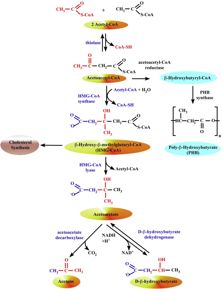
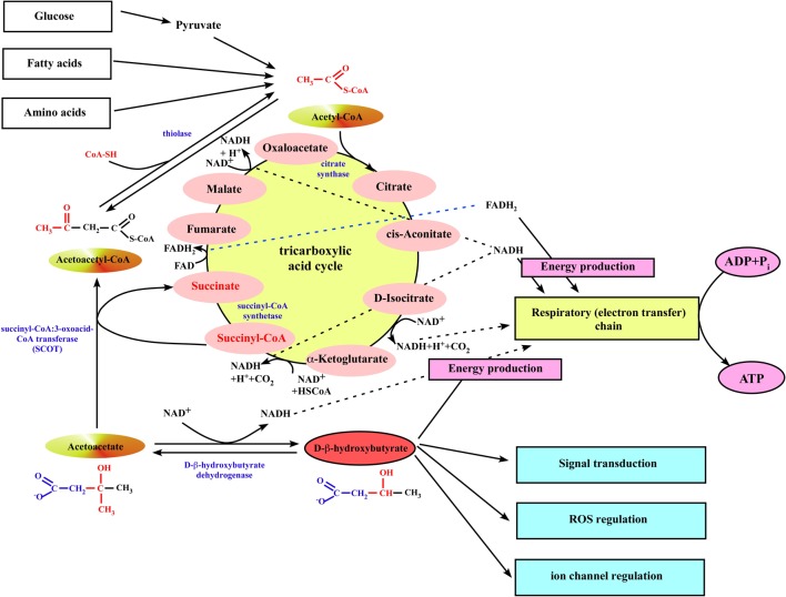
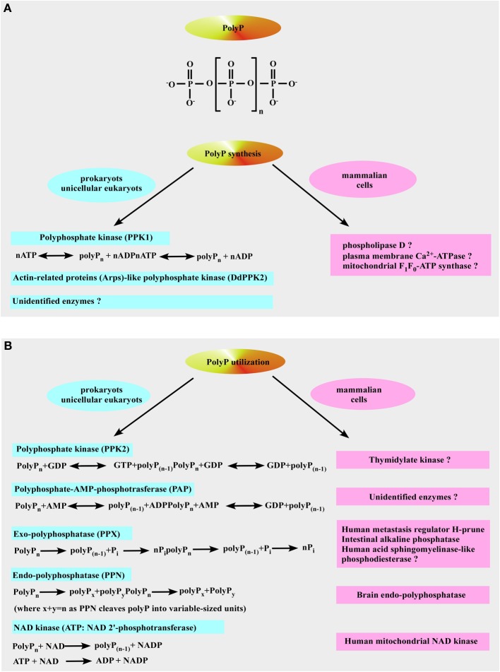
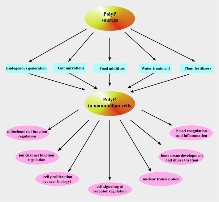
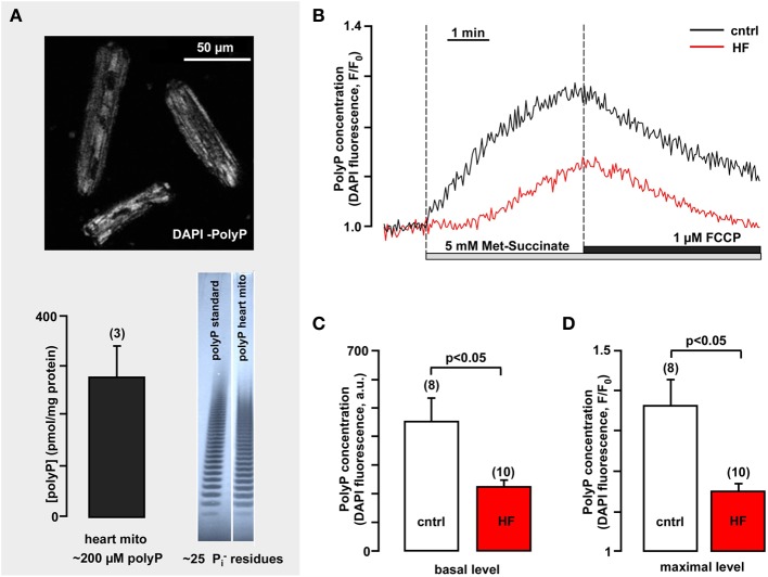
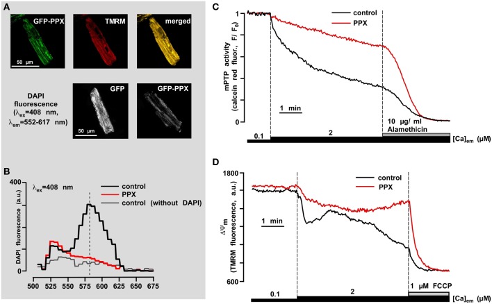
We provide a comprehensive review of the role of β-hydroxybutyrate (β-OHB), its linear polymer poly-β-hydroxybutyrate (PHB), and inorganic polyphosphate (polyP) in mammalian health and disease. β-OHB is a metabolic intermediate that constitutes 70% of ketone bodies produced during ketosis. Although ketosis has been generally considered as an unfavorable pathological state (e.g., diabetic ketoacidosis in type-1 diabetes mellitus), it has been suggested that induction of mild hyperketonemia may have certain therapeutic benefits. β-OHB is synthesized in the liver from acetyl-CoA by β-OHB dehydrogenase and can be used as alternative energy source. Elevated levels of PHB are associated with pathological states. In humans, short-chain, complexed PHB (cPHB) is found in a wide variety of tissues and in atherosclerotic plaques. Plasma cPHB concentrations correlate strongly with atherogenic lipid profiles, and PHB tissue levels are elevated in type-1 diabetic animals. However, little is known about mechanisms of PHB action especially in the heart. In contrast to β-OHB, PHB is a water-insoluble, amphiphilic polymer that has high intrinsic viscosity and salt-solvating properties. cPHB can form non-specific ion channels in planar lipid bilayers and liposomes. PHB can form complexes with polyP and Ca(2+) which increases membrane permeability. The biological roles played by polyP, a ubiquitous phosphate polymer with ATP-like bonds, have been most extensively studied in prokaryotes, however polyP has recently been linked to a variety of functions in mammalian cells, including blood coagulation, regulation of enzyme activity in cancer cells, cell proliferation, apoptosis and mitochondrial ion transport and energy metabolism. Recent evidence suggests that polyP is a potent activator of the mitochondrial permeability transition pore in cardiomyocytes and may represent a hitherto unrecognized key structural and functional component of the mitochondrial membrane system.

Keywords: cardiovascular disease; heart failure; inorganic polyphosphate; mitochondrial permeability transition pore; poly-β-hydroxybutyrate; β-hydroxybutyrate.


Figures

Figure 1
Figure 1
Formation of ketone bodies in liver mitochondria. The synthesis of β-OHB begins with the condensation of two molecules of acetyl-CoA to form acetoacetyl-CoA, the parent of the three ketone bodies, by a ketothiolase enzyme. In prokaryotes, this intermediate is subsequently reduced with NADPH to hydroxybutyryl-CoA by acetoacetyl-CoA reductase, and hydroxybutyryl-CoA may then be polymerized to form PHB by the enzyme PHB synthase. In eukaryotes, 3-hydroxy-3-methylglutaryl-CoA synthase (HMG synthase) catalyzes the condensation of acetoacetyl-CoA with a third acetyl-CoA to form β-hydroxy-β-methylglutaryl-CoA (HMG-CoA). The enzyme HMG-CoA lyase then catalyzes the decomposition of HMG-CoA to form acetoacetate and acetyl-CoA, and acetoacetate is further reduced with NADH by phosphatidylcholine-dependent mitochondrial D-β-hydroxybutyrate dehydrogenase to form β-OHB (Lehninger et al., ; Marks et al., 1992). Acetoacetate is also non-enzymatically decarboxylated to acetone.
Figure 2
Figure 2
Ketone body utilization in mitochondria of extrahepatic organs. When ketone bodies are delivered to the peripheral organs, β-OHB is oxidized back to acetoacetate by the mitochondrial BDH1. Then, acetoacetate gets converted to acetoacetyl-CoA by the mitochondrial enzyme succinyl-CoA:3-oxoacid-CoA transferase (SCOT). The succinyl-CoA:3-oxoacid-CoA transferase uses succinyl-CoA as the CoA donor, forming succinate and acetoacetyl-CoA. This reaction bypasses the succinyl-CoA synthetase step of the TCA cycle, although it does not alter the amount of carbon in the cycle. Next, mitochondrial thiolase cleaves the acetoacetyl-CoA into two molecules of acetyl-CoA, which can generate energy by entering the TCA cycle pathway.
Figure 3
Figure 3
Inorganic polyphosphate structure, formation, and utilization. (A) The upper panel shows the structure of inorganic polyphosphate. The n represents the number of phosphate residues in the polyphosphate chain. It could vary from ten to hundreds of units. The bottom panel is a schematic representation of polyP formation by polyP-generating enzymes in prokaryotes (left) and the putative polyP-generating enzymes in mammalian cells (right). (B) Identified pathways and enzymes for polyP utilization in prokaryotes are shown on the left. Mammalian enzymes which resemble the function of polyP-utilizing enzymes in prokaryotes are shown on the right (see text for more details).
Figure 4
Figure 4
Inorganic polyphosphate sources and functions in mammalian cells. Shown is a schematic representation of the described pathways of polyP occurrence and functions in mammalian cells.
Figure 5
Figure 5
Mitochondrial polyP concentration is highly variable in healthy and heart failure cardiomyocytes and depends on respiratory chain activity. (A) The upper panel image demonstrates polyP detection in freshly isolated rabbit ventricular myocytes using DAPI as a sensor for polyP (λex = 408 nm, λem = 552–617 nm). The bottom panel shows the average amount of polyP in rabbit heart mitochondria (left) and gel images of polyP standard and polyP sample from isolated rabbit mitochondria (right). (B) Original recordings of DAPI fluorescence changes in intact cardiac myocytes stimulated with 5 mM methyl-succinate followed by 5 μM FCCP from control (black) and failing myocytes (red). DAPI fluorescence represents changes in polyP concentration. (C) Average values of basal DAPI fluorescence in control (black) and heart failure (red) myocytes. (D) Average values of maximal DAPI fluorescence after methyl-succinate addition in control (black) and heart failure (red) cells. Modified with permission from Seidlmayer et al. (2012a,b).
Figure 6
Figure 6
PolyP depletion prevents opening of the permeability transition pore induced by mitochondrial Ca2+ overload. (A) The upper panel shows co-localization of GFP-PPX signal with mitochondria. TMRM was used as a mitochondrial signal and the degree of overlay is presented in shades of yellow in the merged image. The bottom panel shows the decrease in DAPI fluorescence in polyP-depleted cells. (B) Fluorescence spectrum of DAPI (5 μM) loaded myocytes expressing control GFP (black), PPX (red), and control GFP cells not loaded with DAPI (gray). (C) Original recordings of mPTP opening using calcein red release from mitochondria of permeabilized control (black) and polyP-depleted (PPX expressing, red) myocytes. After permeabilization cells were exposed to 2 μM Ca2+ and 10 μg/ml alamethicin was added at the end of the experiment to achieve the maximal calcein red release from mitochondria. (D) Original recordings of mitochondrial membrane potential (ΔΨm) with the voltage-sensitive dye TMRM in permeabilized cells upon elevation of the [Ca2+]em from 0.1 to 2 μM, and subsequent addition of 1 μM FCCP in control (black) and polyP-depleted (PPX expressing, red) myocytes. Modified with permission from Seidlmayer et al. (2012b).

Similar articles

Cited by

References

    1. Abramov A. Y., Fraley C., Diao C. T., Winkfein R., Colicos M. A., Duchen M. R., et al. (2007). Targeted polyphosphatase expression alters mitochondrial metabolism and inhibits calcium-dependent cell death. Proc. Natl. Acad. Sci. U.S.A. 104, 18091–18096 10.1073/pnas.0708959104 - DOI - PMC - PubMed
    1. Al-Zaid N. S., Dashti H. M., Mathew T. C., Juggi J. S. (2007). Low carbohydrate ketogenic diet enhances cardiac tolerance to global ischaemia. Acta Cardiol. 62, 381–389 10.2143/AC.62.4.2022282 - DOI - PubMed
    1. Anderson A. J., Dawes E. A. (1990). Occurrence, metabolism, metabolic role, and industrial uses of bacterial polyhydroxyalkanoates. Microbiol. Rev. 54, 450–472 - PMC - PubMed
    1. Aneja P., Dziak R., Cai G. Q., Charles T. C. (2002). Identification of an acetoacetyl coenzyme A synthetase-dependent pathway for utilization of L-(+)-3-hydroxybutyrate in Sinorhizobium meliloti. J. Bacteriol. 184, 1571–1577 10.1128/JB.184.6.1571-1577.2002 - DOI - PMC - PubMed
    1. Arnlov J., Carlsson A. C., Sundstrom J., Ingelsson E., Larsson A., Lind L., et al. (2013). Serum FGF23 and risk of cardiovascular events in relation to mineral metabolism and cardiovascular pathology. Clin. J. Am. Soc. Nephrol. 8, 781–786 10.2215/CJN.09570912 - DOI - PMC - PubMed