. 2014 Jul 22:5:228.
doi: 10.3389/fgene.2014.00228. eCollection 2014.

SCF(SLF)-mediated cytosolic degradation of S-RNase is required for cross-pollen compatibility in S-RNase-based self-incompatibility in Petunia hybrida

Affiliations

SCF(SLF)-mediated cytosolic degradation of S-RNase is required for cross-pollen compatibility in S-RNase-based self-incompatibility in Petunia hybrida

Wei Liu et al. Front Genet. .

Abstract

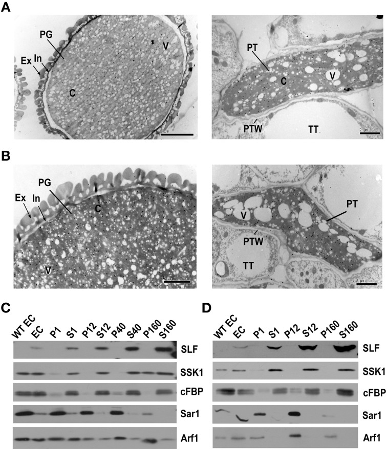
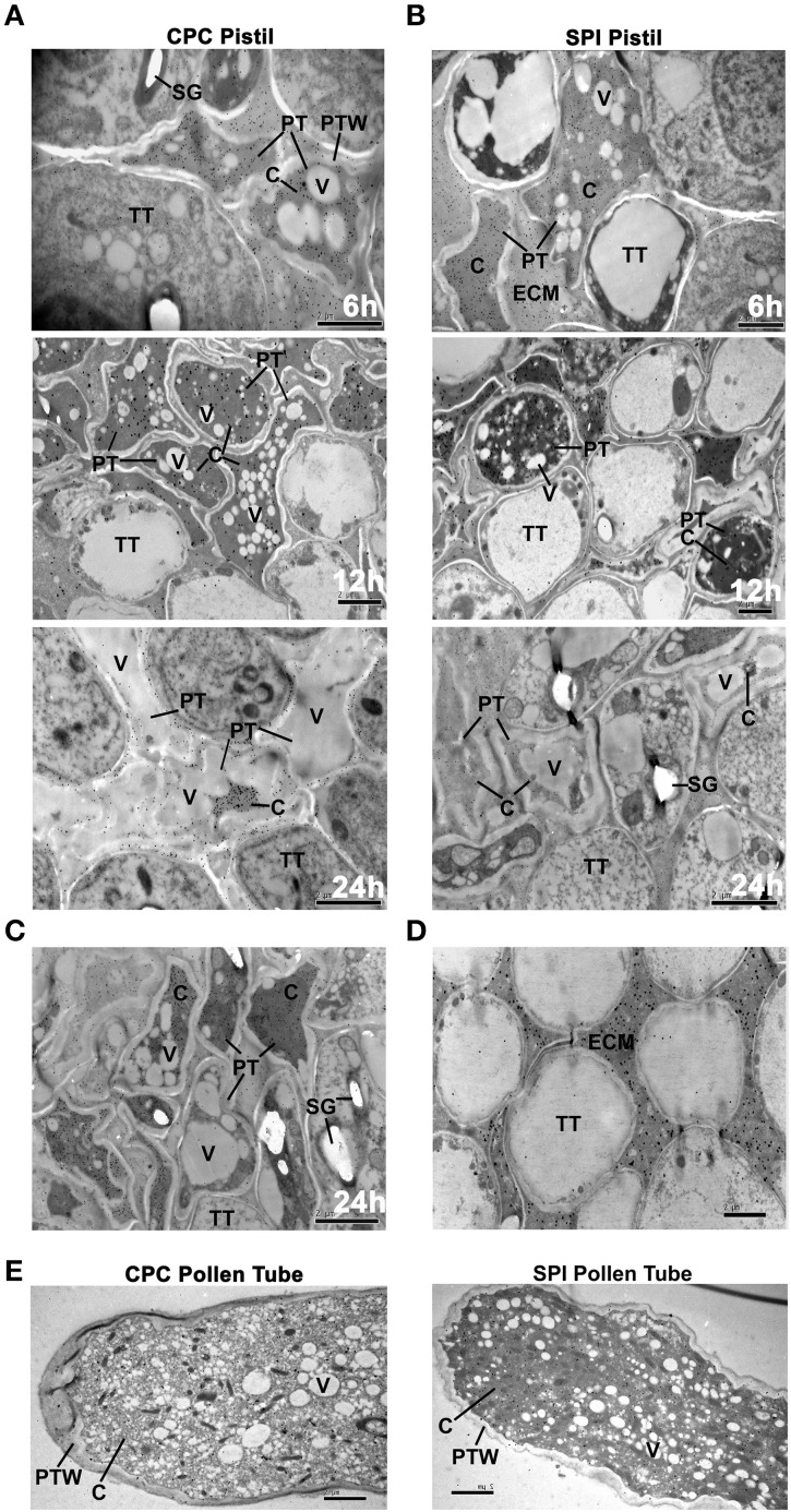
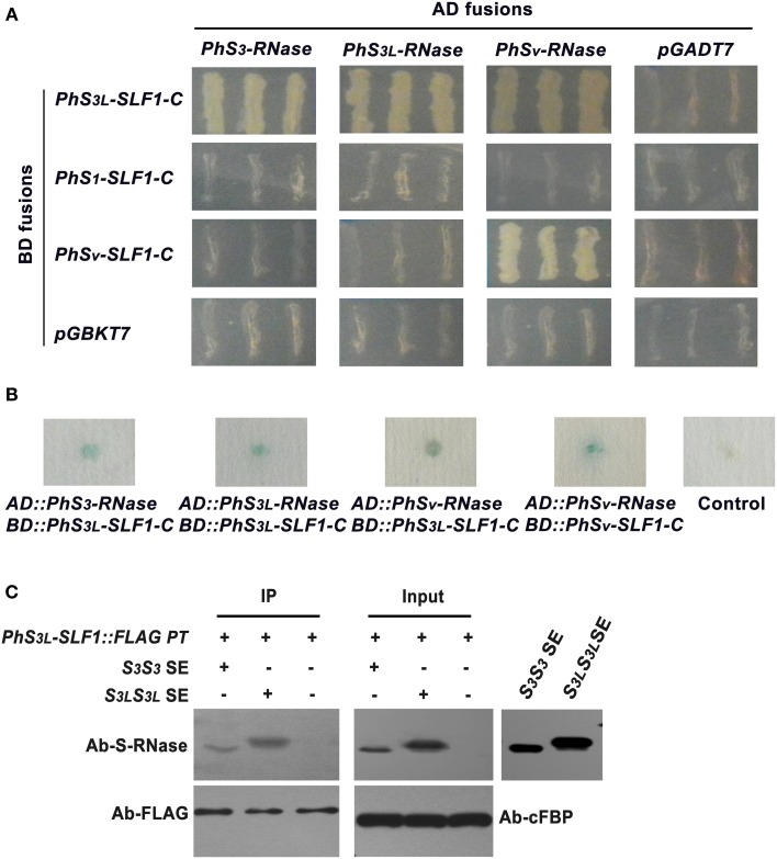
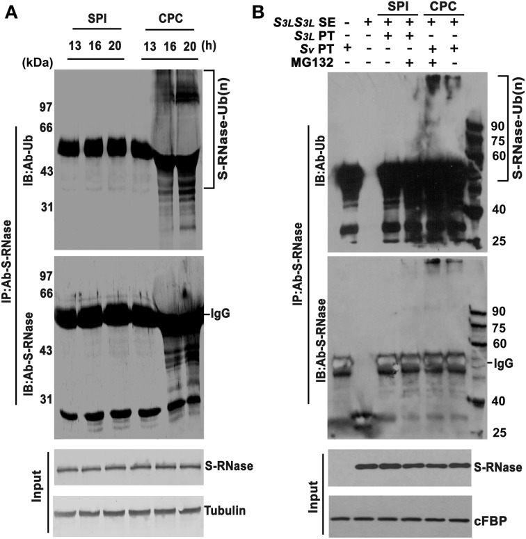
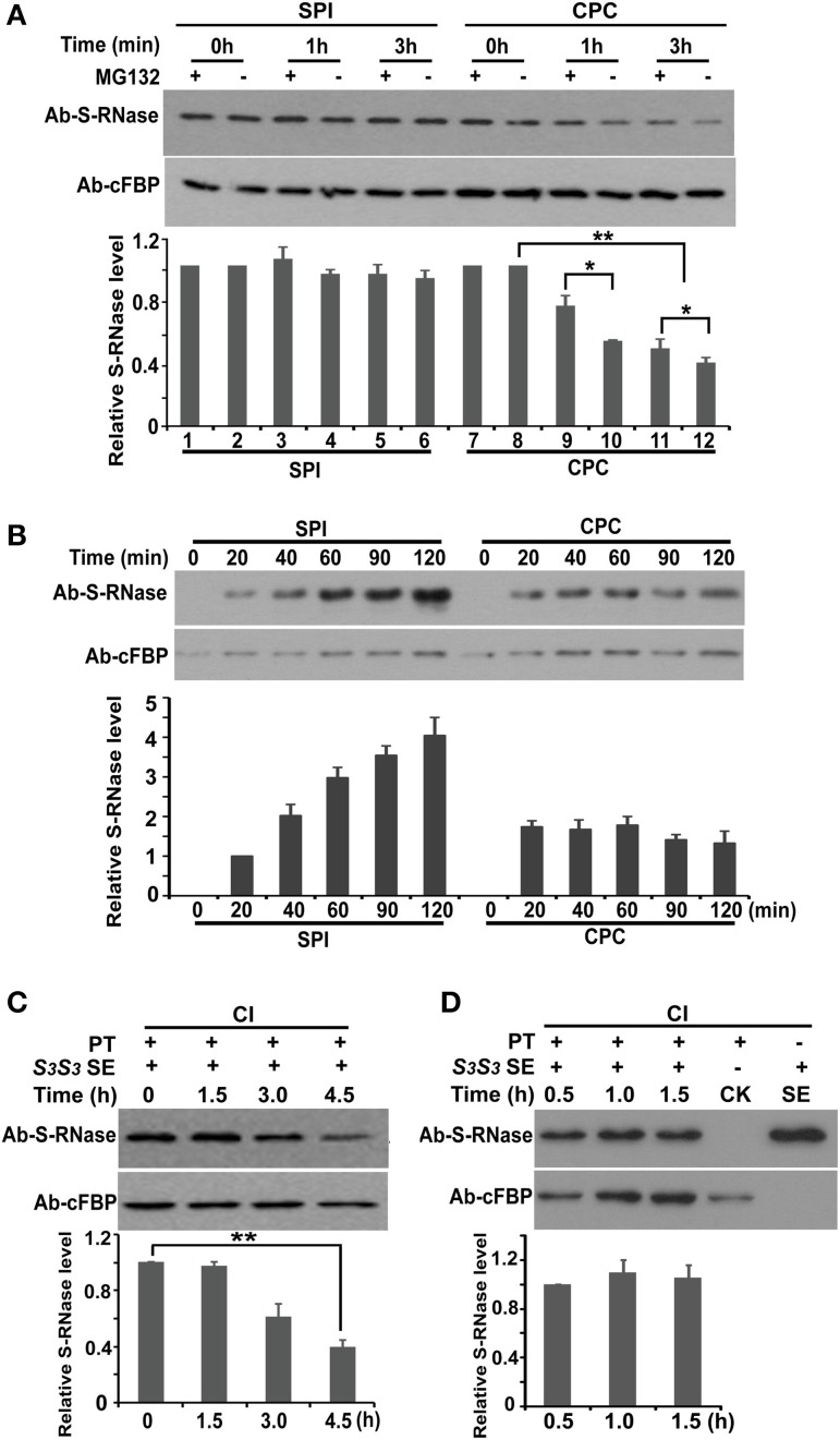
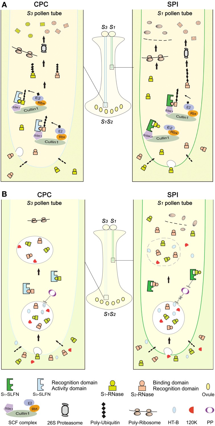
Many flowering plants adopt self-incompatibility (SI) to maintain their genetic diversity. In species of Solanaceae, Plantaginaceae, and Rosaceae, SI is genetically controlled by a single S-locus with multiple haplotypes. The S-locus has been shown to encode S-RNases expressed in pistil and multiple SLF (S-locus F-box) proteins in pollen controlling the female and male specificity of SI, respectively. S-RNases appear to function as a cytotoxin to reject self-pollen. In addition, SLFs have been shown to form SCF (SKP1/Cullin1/F-box) complexes to serve as putative E3 ubiquitin ligase to interact with S-RNases. Previously, two different mechanisms, the S-RNase degradation and the S-RNase compartmentalization, have been proposed as the restriction mechanisms of S-RNase cytotoxicity allowing compatible pollination. In this study, we have provided several lines of evidence in support of the S-RNase degradation mechanism by a combination of cellular, biochemical and molecular biology approaches. First, both immunogold labeling and subcellular fractionation assays showed that two key pollen SI factors, PhS3L-SLF1 and PhSSK1 (SLF-interacting SKP1-like1) from Petunia hybrida, a Solanaceous species, are co-localized in cytosols of both pollen grains and tubes. Second, PhS3L-RNases are mainly detected in the cytosols of both self and non-self-pollen tubes after pollination. Third, we found that PhS-RNases selectively interact with PhSLFs by yeast two-hybrid and co-immunoprecipitation assays. Fourth, S-RNases are specifically degraded in compatible pollen tubes by non-self SLF action. Taken together, our results demonstrate that SCF(SLF-mediated) non-self S-RNase degradation occurs in the cytosol of pollen tube through the ubiquitin/26S proteasome system serving as the major mechanism to neutralize S-RNase cytotoxicity during compatible pollination in P. hybrida.

Keywords: S-RNase localization; SCFSLF; cross-pollen compatibility; self-incompatibility; self-pollen incompatibility; ubiquitin/26S proteasome system.


Figures

Figure 1
Figure 1
PhS3L-SLF1 and PhSSK1 are both located in the cytosol of pollen grains and pollen tubes. (A) Immunogold labeling of PhS3L-SLF1::FLAG in pollen grain (left) and pollen tube (right). (B) Immunogold labeling of PhSSK1 in pollen grain (left) and pollen tube (right). PG, pollen grain; Ex, exine; In, intine (arrow); C, cytosol; V, vacuole; PT, pollen tube; PTW, pollen tube wall; TT, transmitting tract tissue. (C) Western blot detection of PhS3L-SLF1::FLAG (SLF) and PhSSK1 (SSK1) in subcellular fractions of pollen grains. (D) Western blot detection of PhS3L-SLF1::FLAG (SLF) and PhSSK1 (SSK1) in subcellular fractions of in vitro germinated pollen tubes. WT EC and EC denote entire cell homogenates from wild-type and the transgenic pollen grains or pollen tubes, respectively. Pellet fractions (P1, P12, P40, and P160) and supernatant fractions (S1, S12, S40, and S160) are derived from differential centrifugation at 1000, 12,000, 40,000, and 160,000 g, respectively. cFBP, Sar1, and Arf1 are marker antibodies for cytosol, endoplasmic reticulum (ER) and Golgi, respectively.
Figure 2
Figure 2
S-RNases localize to the cytosol of both compatible and incompatible pollen tubes. (A) Immunogold labeling of PhS3L-RNase in cross-sectioned pistils of 6, 12, and 24 h post compatible pollination (CPC), respectively. A zone that is relatively close to the pollen tube tip was observed. (B) Immunogold labeling of PhS3L-RNase in cross-sectioned pistils after 6, 12, and 24 h post incompatible pollination (SPI), respectively. A zone that is relatively close to the pollen tube tip was observed. (C) Immunogold labeling of PhS3L-RNase in cross-sectioned pistils after 24 h post compatible pollination. A zone that is relatively close to the upper one-third pollen tube was observed. (D) A region without pollen tube showing PhS3L-RNase accumulation mainly in ECM of a mature pistil as positive control. (E) Immunogold labeling of PhS3L-RNase in in vitro germinated pollen tube of CPC (Sv) and SPI (S3L), respectively (longitudinal-section). PT, pollen tube; PTW, pollen tube wall; TT, transmitting tract tissue; C, cytosol; V, vacuole; SG, starch granule; ECM, extracellular matrix.
Figure 3
Figure 3
PhSLFs selectively interact with PhS-RNases. (A) Yeast two-hybrid assays between PhSLFs and PhS-RNases. Cells of yeast strain AH109 containing various combinations of bait (BD fusions) and prey (AD fusions) were tested for their growth on selective medium SD/-Ade-His-Leu-Trp. The empty vectors pGBKT7 and pGADT7 were negative controls. (B) The β-galactosidase activity assay was used to further test the interaction of PhSLFs and PhS-RNases. The combination of empty pGBKT7 and pGADT7 was used as a negative control. (C) Co-immunoprecipitation assays between PhS3L-SLF1::FLAG and PhS3-RNase or PhS3L-RNase using in vitro pollen germination system. Untreated pollen tubes were used as negative control (CK). Samples from SPI (treated with S3S3 SE), CPC (treated with S3LS3LSE) and CK pollen tubes were immunoprecipitated by FLAG antibody and detected by PhS-RNase antibody and FLAG antibody, respectively. Challenged but not immmunoprecipitated SPI, CPC, and CK pollen tube samples were loaded as input. cFBP was detected as input loading control. PT, pollen tube; SE, style extract.
Figure 4
Figure 4
S-RNases are polyubiquitinated in compatible pollen tubes. (A) PhS-RNases are polyubiquitinated in pollen tubes of compatible pollination. Pollen tubes were dissected from styles after 13, 16, and 20 h post-pollination. Samples extracted and immunoprecipitated by PhS-RNase antibody and detected by ubiquitin (Ub) antibody and PhS-RNase antibody. PhS-RNase and tubulin were detected as input and loading control. (B) PhS-RNases are polyubiquitinated in CPC response in in vitro pollen germination system. In vitro germinated S3L (SPI) and Sv (CPC) pollen tubes challenged by S3LS3L style extract were subjected to MG132 treatment (+) or not (−). Samples of SPI, CPC, and CK were immunoprecipitated by PhS-RNase antibody and detected by ubiquitin (Ub) antibody and PhS-RNase antibody, respectively. Style extracts and immunoprecipitation of the untreated pollen tubes samples were used as negative controls. Challenged but not immmunoprecipitated pollen tube samples were loaded as input. PhS3L-RNase and cFBP were detected as input and loading control. S-RNase-Ub (n) denotes polyubiquitinated S-RNase. IgG indicates heavy chain. Molecular weights in kilodalton (kDa) are shown on right or left side of the blots.
Figure 5
Figure 5
SCFSLF directly mediates non-self S-RNase degradation by the 26S proteasome pathway. (A) Time course analysis of PhS3L-RNase levels in pulse challenged SPI (S3L) and CPC (Sv) pollen tubes in conjunction with (+) or without (−) MG132 treatment. (B) Time course analysis of PhS3L-RNase accumulations in continuously challenged SPI (S3L) and CPC (Sv) pollen tubes. In vitro germinated S3L and Sv pollen tubes were continuously treated with S3LS3L style extract. PhS3L-RNase levels in pollen tubes were monitored by western blot analysis. cFBP was detected as loading control. (C) Time course analysis of PhS3-RNase levels in pulse challenged pollen tubes with stylar extracts. The pollen tubes were derived from the transgenic plants exhibiting competitive interaction (CI) between the transgene PhS3L-SLF1 and S3 haplotype. PhS3-RNase was detected by western blot to monitor its dynamics in S3/PhS3L-SLF1 pollen tubes. (D) Time course analysis of PhS3-RNase levels in continuously challenged pollen tubes with stylar extracts. The transgenic S3/PhS3L-SLF1 pollen tubes were continuously treated with S3S3 style extract and monitored for S3-RNase level through the time course. CK indicates pollen tubes before treatment. cFBP was detected as loading control. PT, S3/PhS3L-SLF1 pollen tube; SE, style extract. Column charts in both (A–D) show quantitative S-RNases levels determined by Quantity One software using three replicates. T tests were done between the designated lanes. Single and double asterisks denote significant and extremely significant difference, respectively.
Figure 6
Figure 6
Models of S-RNase-based self-incompatibility. (A) S-RNase degradation model in Petunia. In CPC response, cross S3 pollen lands on the stigma, germinates and grows into an S1S2 style. Both S1- and S2-RNases enter the S3 pollen tube by endocytosis or an unknown pathway (dotted arrows). Subsequently, the hypothetical binding domains of S1- and S2-RNases interact with a hypothetical activity domain of the pollen S (i.e., SLF in Solanaceae and Plantaginaceae) in the cytosol of the pollen tube. Then, one or more SLF (SLFN) in S3 pollen tube form functional SCFS3-SLFN complexes to tag S1- and S2-RNases with a polyubiquitin chain, which are subsequently degraded by the 26S proteasome. Thus, ribosomal RNAs escape from degradation by S-RNases in cross-pollen tubes, resulting in normal pollen tube growth. In SPI, during which self S1 pollen lands on the S1S2 style, both S1- and S2-RNases enter the S1 pollen tube. Similar to CPC response, non-self S2-RNases bind to a hypothetical activity domain of the pollen S in the cytosol of the pollen tube. Then, one or more SLF (SLFN) in S1 pollen tube form SCFS1-SLFN complexes to tag S2-RNase with a polyubiquitin chain, resulting in its degradation by the 26S proteasome. In contrast, the recognition domain of self S1-RNase binds to a hypothetical recognition domain of SLF resulting in the formation of a non-functional SCFS1-SLFN complex, thus self S-RNase escapes degradation and acts as a cytotoxin to inhibit the pollen tube growth. (B) S-RNase compartmentalization model in Nicotiana. S-RNases, 120K and HT-B all enter the pollen tubes. In CPC response, HT-B degradation possibly occurs by a hypothetical pollen protein (PP), thus S-RNase is sorted to a vacuolar compartment. In SPI, the interaction between S-RNase and SLF has been postulated to stabilize HT-B, perhaps by inhibiting the PP action. Subsequently, the vacuolar compartment breaks down, releasing S-RNase and acts as a cytotoxin to inhibit the pollen tube growth.

Similar articles

Cited by

References

    1. Agrawal G. K., Bourguignon J., Rolland N., Ephritikhine G., Ferro M., Jaquinod M., et al. (2011). Plant organelle proteomics: collaborating for optimal cell function. Mass Spectrom. Rev. 30, 772–853 10.1002/mas.20301 - DOI - PubMed
    1. Anderson M. A., McFadden G. I., Bernatzky R., Atkinson A., Orpin T., Dedman H., et al. (1989). Sequence variability of three alleles of the self-incompatibility gene of Nicotiana alata. Plant Cell 1, 483–491 10.1105/tpc.1.5.483 - DOI - PMC - PubMed
    1. Arrese E. L., Gazard J. L., Flowers M. T., Soulages J. L., Wells M. A. (2001). Diacylglycerol transport in the insect fat body: evidence of involvement of lipid droplets and the cytosolic fraction. Lipid Res. 42, 225–234 - PubMed
    1. Bai C., Sen P., Hofmann K., Ma L., Goebl M., Harper J. W., et al. (1996). SKP1 connects cell cycle regulators to the ubiquitin proteolysis machinery through a novel motif, the F-box. Cell 86, 263–274 10.1016/S0092-8674(00)80098-7 - DOI - PubMed
    1. Busot G. Y., McClure B., Ibarra-Sánchez C. P., Jiménez-Durán K., Vázquez-Santana S., Cruz-García F. (2008). Pollination in Nicotiana alata stimulates synthesis and transfer to the stigmatic surface of NaStEP, a vacuolar Kunitz proteinase inhibitor homologue. J. Exp. Bot. 59, 3187–3201 10.1093/jxb/ern175 - DOI - PMC - PubMed

LinkOut - more resources