

Varicocele Time-dependently Affects DNA Integrity of Sperm Cells: Evidence for Lower In vitro Fertilization Rate in Varicocele-positive Rats
- PMID: 25101162
- PMCID: PMC4122833
Varicocele Time-dependently Affects DNA Integrity of Sperm Cells: Evidence for Lower In vitro Fertilization Rate in Varicocele-positive Rats
Abstract
Background: We designed this study to clarify how varicocele can time-dependently affect sperm morphological parameters and DNA integrity. In this study, we intend to estimate the effect of various periods of varicocele on the in vitro fertilization (IVF) rate in rats.
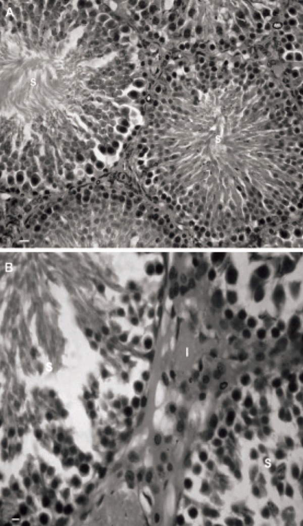
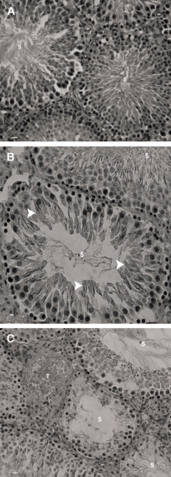
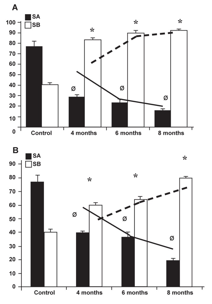
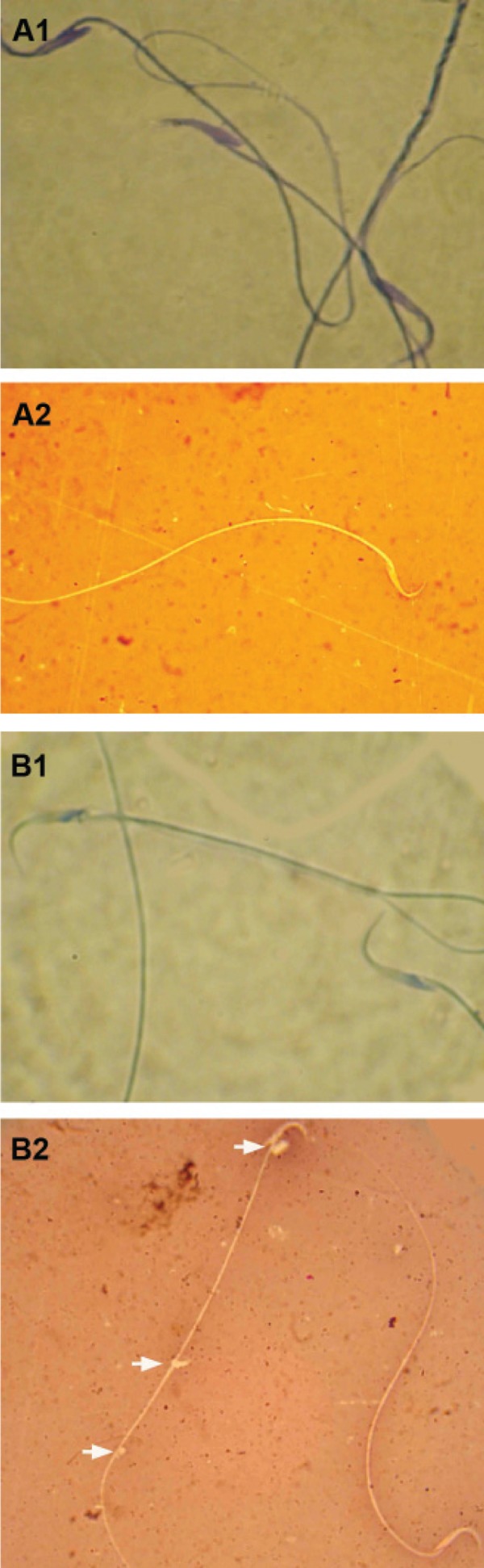
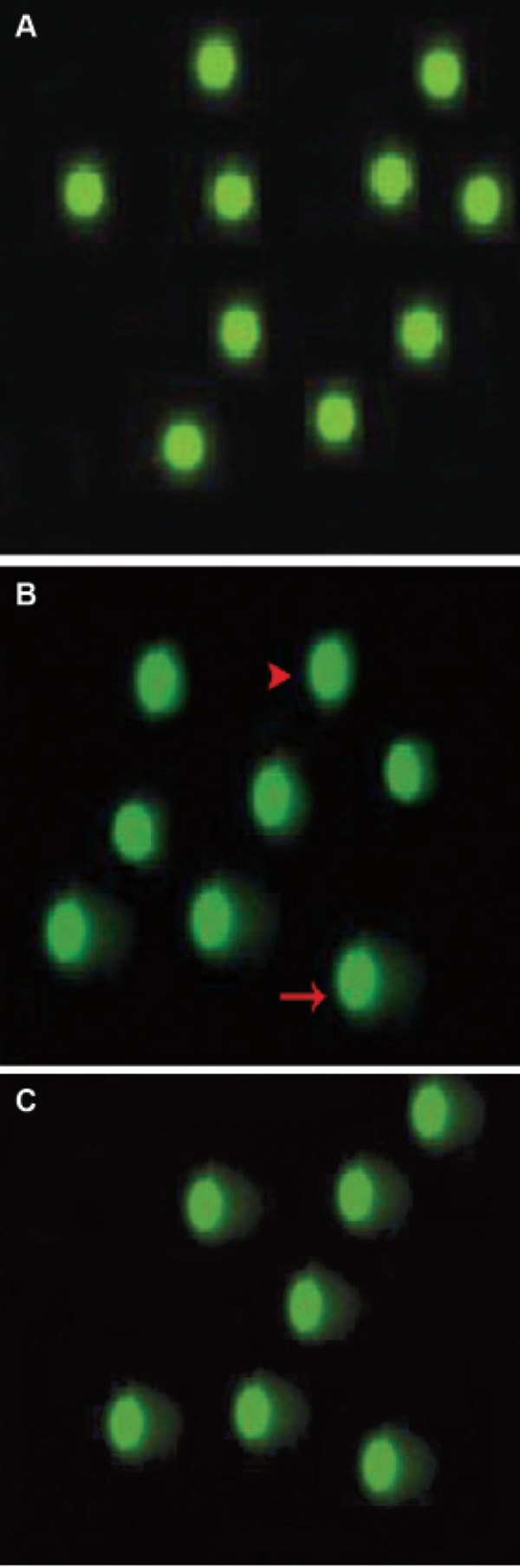
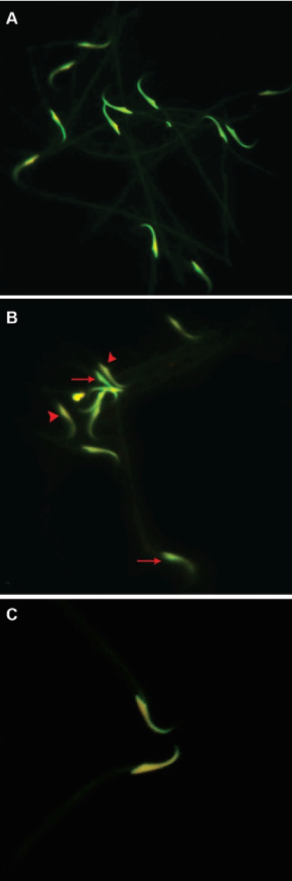
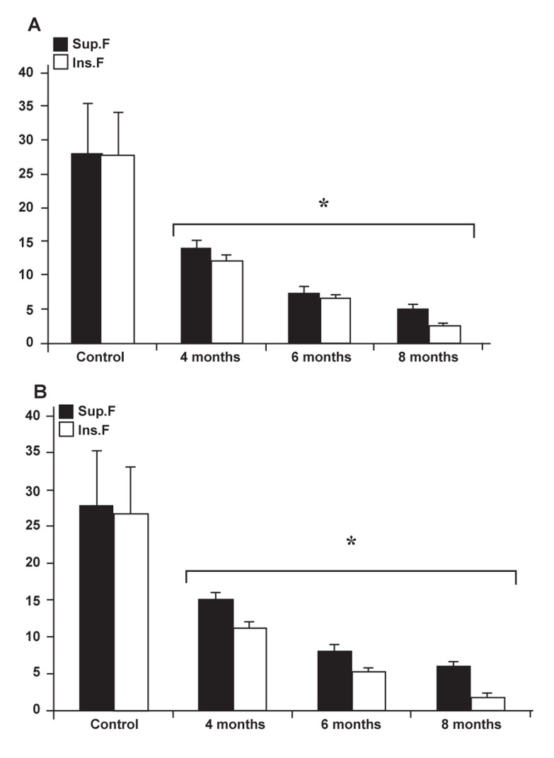
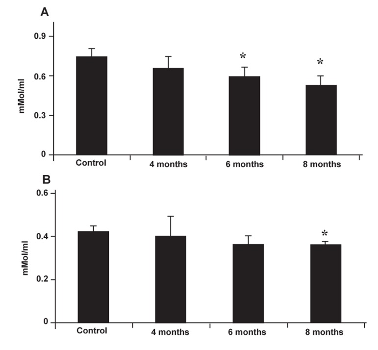
Materials and methods: In this experimental study, left varicocele were induced as the test group (n=18) which was further sub-divided into three groups based on the study termination time (4, 6 and 8 months after varicocele induction). The control-sham group (n=6) consisted of rats who received no treatment. Repopulation index (RI), tubular differentiation index (TDI), sperm viability and motility, morphological maturity, chromatin integrity and ability to undergo IVF were assessed. In addition, the potential impact of varicocele on serum total antioxidant capacity (TAOC) and total thiol molecules (TTM) were examined.
Results: Histological results showed that varicocele negatively influenced TDI and RI. All sperm morphological parameters were lower than those in the control-sham group. DNA damage was severely and time-dependently substantiated in all test groups. Varicocele significantly reduced the ability of sperm derived from varicocele rats to undergo IVF. Serum TAOC and TTM levels reduced in a time-dependent manner. Right testes varicocele-induced rats showed remarkably less damaged profile for all investigated parameters compared to the left testes varicocele.
Conclusion: Our data suggested that experimentally induced varicocele negatively impacted sperm maturation and chromatin integrity in a time-dependent manner. This consequently caused a remarkable reduction in IVF ability. The detrimental effect of varicocele may be attributed to the significant reduction of antioxidant capacity of the serum.
Keywords: DNA Damage; In vitro Fertilization; Oxidative Stress; Varicocele.
Figures
Similar articles
-
Silymarin protects from varicocele-induced damages in testis and improves sperm quality: evidence for E2f1 involvement.Syst Biol Reprod Med. 2013 Oct;59(5):270-80. doi: 10.3109/19396368.2013.794253. Epub 2013 May 10. Syst Biol Reprod Med. 2013. PMID: 23659554
-
Evaluation of Oxidative Stress in Testis and Sperm of Rat Following Induced Varicocele.Urol J. 2019 Jun 17;16(3):300-306. doi: 10.22037/uj.v0i0.4740. Urol J. 2019. PMID: 30471074
-
Tracing of zinc and iron in experimentally induced varicocele: correlation with oxidative, nitrosative and carbonyl stress.Andrologia. 2017 Aug;49(6). doi: 10.1111/and.12687. Epub 2016 Sep 29. Andrologia. 2017. PMID: 27682184
-
Varicocele-Induced Infertility in Animal Models.Int J Fertil Steril. 2015 Jul-Sep;9(2):141-9. doi: 10.22074/ijfs.2015.4234. Epub 2015 Jul 27. Int J Fertil Steril. 2015. PMID: 26246871 Free PMC article. Review.
-
Varicocele management in the era of in vitro fertilization/intracytoplasmic sperm injection.Asian J Androl. 2016 May-Jun;18(3):343-8. doi: 10.4103/1008-682X.178482. Asian J Androl. 2016. PMID: 27030086 Free PMC article. Review.
Cited by
-
Oxidative Stress and Cell Cycle Arrest in Seminiferous Tubules Nearby Varicose Vessels: New Perspectives from Experimental Varicocele.Reprod Sci. 2023 Aug;30(8):2401-2415. doi: 10.1007/s43032-023-01200-4. Epub 2023 Feb 23. Reprod Sci. 2023. PMID: 36821035
-
Effect of purine nucleoside analogue-acyclovir on the sperm parameters and testosterone production in rats.Int J Fertil Steril. 2013 Apr;7(1):49-56. Epub 2013 Mar 6. Int J Fertil Steril. 2013. PMID: 24520464 Free PMC article.
-
Molecular Remodeling of the Sperm Proteome Following Varicocele Sclero-Embolization: Implications for Semen Quality Improvement.Proteomes. 2025 Jul 15;13(3):34. doi: 10.3390/proteomes13030034. Proteomes. 2025. PMID: 40700278 Free PMC article.
-
Silymarin and celecoxib ameliorate experimental varicocele-induced pathogenesis: evidences for oxidative stress and inflammation inhibition.Int Urol Nephrol. 2018 Jun;50(6):1039-1052. doi: 10.1007/s11255-018-1862-5. Epub 2018 Apr 5. Int Urol Nephrol. 2018. PMID: 29623501
-
Semen parameters in men with varicocele: DNA fragmentation, chromatin packaging, mitochondrial membrane potential, and apoptosis.JBRA Assist Reprod. 2017 Dec 1;21(4):295-301. doi: 10.5935/1518-0557.20170053. JBRA Assist Reprod. 2017. PMID: 29068181 Free PMC article.
References
-
- French DB, Desai NR, Agarwal A. Varicocele repair: does it still have a role in infertility treatment? Curr Opin Obstet Gynecol. 2008;20(3):269–274. - PubMed
-
- Naughton CK, Nangia AK, Agarwal A. Pathophysiology of varicoceles in male infertility. Hum Reprod Update. 2001;7(5):473–481. - PubMed
-
- Witt MA, Lipshultz LI. Varicocele: a progressive or static lesion? Urology. 1993;42(5):541–543. - PubMed
-
- Kamal KM, Jarvi K, Zini A. Microsurgical varicocelectomy in the era of assisted reproductive technology: in influence of initial semen quality on pregnancy rates. Fertil Steril. 2001;75(5):1013–1016. - PubMed
-
- Skoog SJ, Roberts KP, Goldstein M, Pryor JL. The adolescent varicocele: What’s new with an old problem in young patients? Pediatrics. 1997;100(1):112–121. - PubMed
LinkOut - more resources
Full Text Sources