. 2014 Aug 1:6:16.
doi: 10.1186/2045-824X-6-16. eCollection 2014.

VEGFR-1 blockade disrupts peri-implantation decidual angiogenesis and macrophage recruitment

Affiliations

VEGFR-1 blockade disrupts peri-implantation decidual angiogenesis and macrophage recruitment

Nataki C Douglas et al. Vasc Cell. .

Abstract

Background: Angiogenesis and macrophage recruitment to the uterus are key features of uterine decidualization; the progesterone-mediated uterine changes that allow for embryo implantation and initiation of pregnancy. In the current study, we characterized the expression of vascular endothelial growth factor receptor-1 (VEGFR-1) in macrophages and endothelial cells of the peri-implantation uterus and determined if VEGFR-1 function is required for decidual angiogenesis, macrophage recruitment, and/or the establishment of pregnancy.

Methods: Expression of VEGFR-1 in uterine endothelial cells and macrophages was determined with immunohistochemistry. To assess the effect of continuous VEGFR-1 blockade, adult female mice were given VEGFR-1 blocking antibody, MF-1, every 3 days for 18 days. After 6 doses, females were mated and a final dose of MF-1 was given on embryonic day 3.5. Endothelial cells and macrophages were quantified on embryonic day 7.5. Pregnancy was analyzed on embryonic days 7.5 and 10.5.

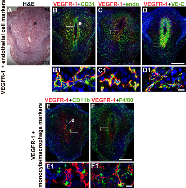
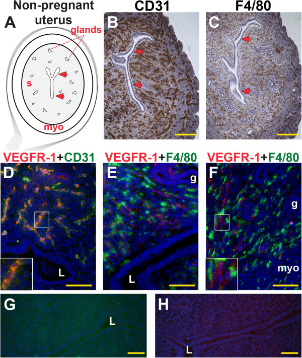
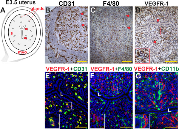
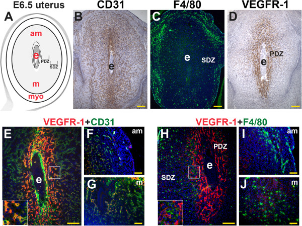
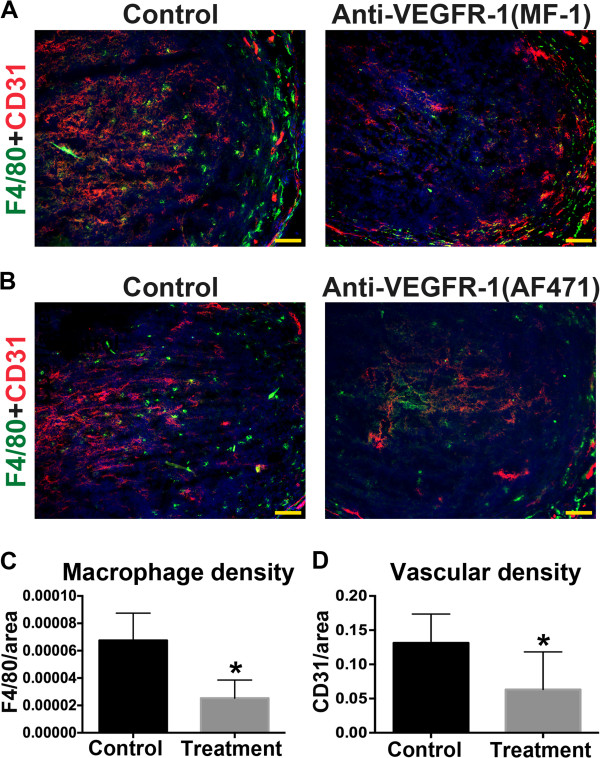
Results: F4/80(+) macrophages are observed throughout the stroma and are abundant adjacent to the endometrial lumen and glands prior to embryo implantation and scatter throughout the decidua post implantation. VEGFR-1 expression is restricted to the uterine endothelial cells. F4/80(+) macrophages were often found adjacent to VEGFR-1(+) endothelial cells in the primary decidual zone. Continuous VEGFR-1 blockade correlates with a significant reduction in decidual vascular and macrophage density, but does not affect embryo implantation or maintenance of pregnancy up to embryonic day 10.5.

Conclusions: We found that VEGFR-1 functions in both decidual angiogenesis and macrophage recruitment to the implantation site during pregnancy. VEGFR-1 is expressed by endothelial cells, however blocking VEGFR-1 function in endothelial cells results in reduced macrophage recruitment to the uterus. VEGFR-1 blockade did not compromise the establishment and/or maintenance of pregnancy.

Keywords: Angiogenesis; Decidua; Endothelial cells; Implantation; Macrophages; Uterus; VEGFR-1.


Figures

Figure 1
Figure 1
VEGFR-1 expression in endothelial cells and macrophages in the post-implantation uterus. H&E and double staining IF were performed on E6.5 frontal uterine sections. (A) H&E of a post-implantation mouse uterus showing the embryo (e), anti-mesometrial (am) and mesometrial (m) areas. (B-F) VEGFR-1+ cells (red) are observed in the decidua, with abundant expression in the primary decidual zone surrounding the implanted embryo. (B) Double-staining for VEGFR-1+ (red) and CD31+ (green) cells demonstrates expression of VEGFR-1 in a subset of CD31+ ECs. (C) Double-staining for VEGFR-1+ (red) and endomucin+ (green) cells demonstrates expression of VEGFR-1 in a subset of endomucin+ ECs. (D) Double-staining for VEGFR-1+ (red) and VE-cadherin+ (green) cells demonstrates expression of VEGFR-1 in VE-cadherin+ ECs. (E) Double-staining for VEGFR-1+ (red) and CD11b+ (green) cells demonstrates that VEGFR-1 is not expressed in CD11b+ monocytes. (F) Double-staining for VEGFR-1+ (red) and F4/80+ (green) cells demonstrates that VEGFR-1 is not expressed in F4/80+ macrophages. VEGFR-1+ cells are adjacent to CD11b+ monocytes and F4/80+ macrophages. White boxes in (B-F) indicate areas of the uteri magnified below (B1-F1). (A-F) Scale bar = 500 μm. (B1-F1) Scale bar = 20 μm.
Figure 2
Figure 2
VEGFR-1, CD31 and F4/80 expression in the non-pregnant uterus. IHC and double staining IF were performed on non-pregnant uterine cross-sections. (A) Schematic representation of a non-pregnant mouse uterus showing lumen (arrowheads), endometrial glands, stroma (s), and myometrium (myo). (B) ECs, detected by CD31 staining (brown), are observed throughout the stroma and myometrium. (C) Macrophages, detected by F4/80 staining (brown), are observed throughout the stroma and myometrium. (D) Double-staining for VEGFR-1+ (red) and CD31+ (green) demonstrates expression of VEGFR-1 in a subset of CD31+ ECs throughout the stroma. (E, F) VEGFR-1+ cells (red) and F4/80+ macrophages (green) are distributed throughout the stroma. VEGFR-1 and F4/80 co-expression is not observed. The inset (F) highlights contact of adjacent VEGFR-1+ cells and F4/80+ macrophages. (G, H) IF controls with secondary antibodies alone show no specific staining. L, lumen; g, endometrial gland. (B, C) Scale bar = 100 μm. (D-H) Scale bar = 50 μm.
Figure 3
Figure 3
VEGFR-1, CD31, F4/80, and CD11b expression in the pre-implantation mouse uterus. IHC and double staining IF were performed on E3.5 uterine cross-sections. (A) Schematic representation of an E3.5 mouse uterus showing lumen (arrowheads), glands, stroma (s), and myometrium (myo). (B) ECs, detected by CD31 staining (brown), are observed throughout the stroma and myometrium, similar to the non-pregnant state. (C) Macrophages, detected by F4/80 staining (brown), are observed throughout the stroma and are abundant adjacent to the lumen and glands at E3.5. (D) VEGFR-1+ cells (brown), are distributed throughout the stroma and cell associated VEGFR-1 expression highlighted in the inset. (E) Double staining for VEGFR-1 (red) and CD31 (green) demonstrates expression of VEGFR-1 on CD31+ ECs throughout the stroma. (F) VEGFR-1+ cells (red) and F4/80+ macrophages (green) are distributed throughout the stroma. VEGFR-1 and F4/80 co-expression is not observed. (G) VEGFR-1+ cells (red) and CD11b+ monocytes (green) are distributed throughout the stroma. VEGFR-1 and CD11b co-expression is not observed. L, lumen. Scale bars B, C = 100 μm. Scale bars D – F = 50 μm.
Figure 4
Figure 4
VEGFR-1, CD31, and F4/80 in the post-implantation mouse uterus. IHC and double staining IF were performed on E6.5 frontal uterine sections. (A) Schematic representation of an E6.5 mouse uterus showing the embryo (e), anti-mesometrial (am) and mesometrial (m) areas, and myometrium (myo). (B) CD31+ ECs (brown) are abundant in the decidua around the implanted embryo and the myometrium. (C) A majority of F4/80+ macrophages (green) are observed in the secondary decidual zone (SDZ) and myometrium. (D) A majority of VEGFR-1+ cells (brown) are observed in the primary decidual zone (PDZ) directly adjacent to the implanted embryo. (E-F) Double staining of VEGFR-1 (red) and CD31+ (green) demonstrates (E) co-expression in the ECs within the PDZ (inset), (F, G) and minimal co-expression of VEGFR-1 and CD31 in the abundant ECs in the SDZ of the anti-mesometrial and mesometrial poles. (H) Double staining for VEGFR-1 (red) and F4/80+ (green) shows VEGFR-1+ cells adjacent to F4/80+ macrophages in the PDZ (inset). (I, J) F4/80+ macrophages are abundant in the SDZ of the anti-mesometrial pole and mesometrial poles, where VEGFR-1 expression is low to absent. (B-D, H) Scale bars = 100 μm. (F, G, I, J) Scale bars = 50 μm.
Figure 5
Figure 5
VEGFR-1 blockade reduces peri-implantation macrophage and vascular density at E7.5. (A, B) Double staining for F4/80 (green) and CD31 (red) in E7.5 uterine frontal sections from mice treated with VEGFR-1 blocking antibodies (MF-1 or AF471) or from control mice. Reduced expression of F4/80 and CD31 is observed in treatment groups. Scale bars = 50 μm. (C, D) F4/80+ or CD31+ signal was determined and normalized by total decidual area. F4/80+ macrophage and CD31+ EC density is significantly reduced in uteri of mice that received MF-1 compared to controls.

References

    1. Dey SK, Lim H, Das SK, Reese J, Paria BC, Daikoku T, Wang H. Molecular cues to implantation. Endocr Rev. 2004;25:341–373. - PubMed
    1. Cha J, Sun X, Dey SK. Mechanisms of implantation: strategies for successful pregnancy. Nat Med. 2012;18:1754–1767. - PMC - PubMed
    1. Wang H, Dey SK. Roadmap to embryo implantation: clues from mouse models. Nat Rev Genet. 2006;7:185–199. - PubMed
    1. Kabawat SE, Mostoufi-Zadeh M, Driscoll SG, Bhan AK. Implantation site in normal pregnancy. A study with monoclonal antibodies. Am J Pathol. 1985;118:76–84. - PMC - PubMed
    1. Fest S, Aldo PB, Abrahams VM, Visintin I, Alvero A, Chen R, Chavez SL, Romero R, Mor G. Trophoblast-macrophage interactions: a regulatory network for the protection of pregnancy. Am J Reprod Immunol. 2007;57:55–66. - PubMed

LinkOut - more resources