Review
. 2014 Mar 25:2014:928461.
doi: 10.1155/2014/928461. eCollection 2014.

Role of Th17 Cells in the Pathogenesis of Human IBD

Affiliations
Review

Role of Th17 Cells in the Pathogenesis of Human IBD

Julio Gálvez. ISRN Inflamm. .

Abstract

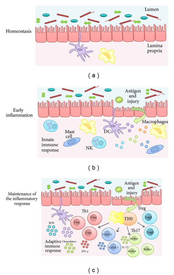
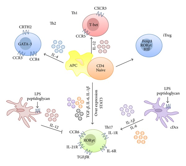
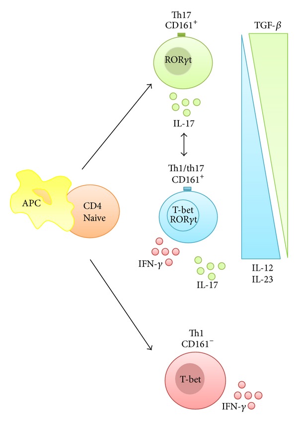
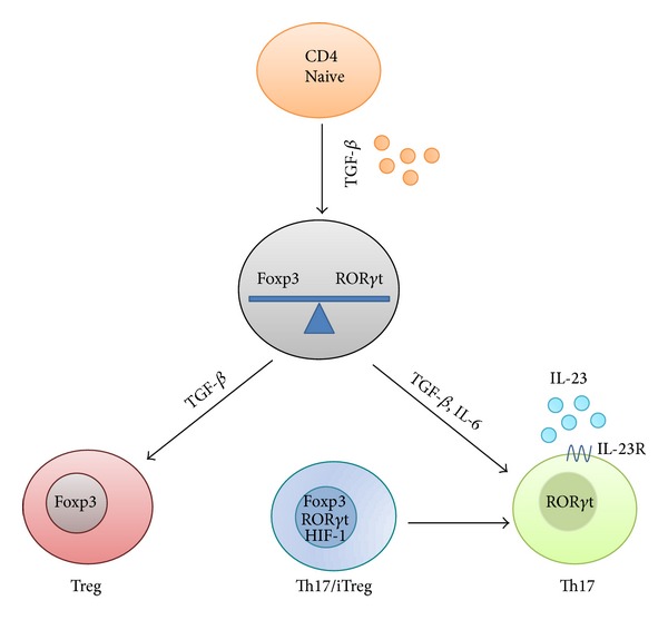
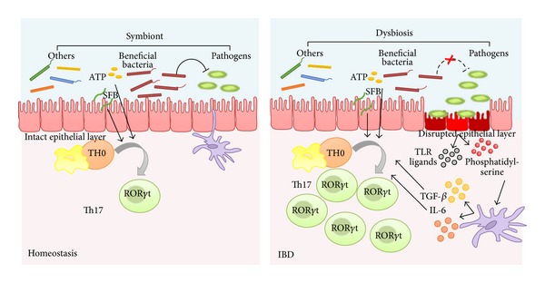
The gastrointestinal tract plays a central role in immune system, being able to mount efficient immune responses against pathogens, keeping the homeostasis of the human gut. However, conditions like Crohn's disease (CD) or ulcerative colitis (UC), the main forms of inflammatory bowel diseases (IBD), are related to an excessive and uncontrolled immune response against normal microbiota, through the activation of CD4(+) T helper (Th) cells. Classically, IBD was thought to be primarily mediated by Th1 cells in CD or Th2 cells in UC, but it is now known that Th17 cells and their related cytokines are crucial mediators in both conditions. Th17 cells massively infiltrate the inflamed intestine of IBD patients, where they produce interleukin- (IL-) 17A and other cytokines, triggering and amplifying the inflammatory process. However, these cells show functional plasticity, and they can be converted into either IFN- γ producing Th1 cells or regulatory T cells. This review will summarize the current knowledge regarding the regulation and functional role of Th17 cells in the gut. Deeper insights into their plasticity in inflammatory conditions will contribute to advancing our understanding of the mechanisms that regulate mucosal homeostasis and inflammation in the gut, promoting the design of novel therapeutic approaches for IBD.


Figures

Figure 1
Figure 1
Physiopathology of IBD. (a) The intestine comprises the major single epithelial interface in the body, which is populated by the greatest number and diversity of resident microbes. The intestinal immune system therefore encounters more antigens than any other part of the body and it must discriminate between invasive organisms and harmless antigens, such as food, proteins, and commensal bacteria. The intestinal homeostasis depends on the dynamic crosstalk between the microbiota, the intestinal epithelial cells, and the resident immune cells. (b) Several mechanisms are involved in the regulation of the intestinal homeostasis. The breakdown of this balance triggers the chronic inflammatory process found in inflammatory bowel disease. During early inflammation, foreign antigens activate the different innate immune cells located in the intestine, including natural killer cells, mast cells, neutrophils, macrophages, and dendritic cells. (c) A maintained inflammatory reaction promotes the activation of the adaptive immune response. Abnormally activated effector CD4+ T helper (Th) cells synthetize and release different inflammatory mediators that generate the vicious circle of inflammation that leads to chronic tissue injury and epithelial damage.
Figure 2
Figure 2
T-cell differentiation into Th17 cells. Naive CD4+ T cells, upon contact with antigen-presenting cells (APCs), have the potential to differentiate into Th1, Th2, Th17, and induced regulatory T cells (iTreg), a process controlled by the effector cytokines produced by APCs. Th17 cells are originated from a small subset of naive T cells that express CD161 and cytokine receptors IL-6R, TGFβR, IL-23R, IL-21R, and IL-1R, being their polarization into Th17 cells promoted by the combined activity of these cytokines. These cytokines mediate their effects by promoting or repressing the expression of different transcription factors, in particular STAT3 and RORγt, which are critical for Th17 differentiation and development. It is of great interest to discern the nature of the APCs capable of inducing Th17 responses and the corresponding stimuli involved in this. Monocytes and circulating conventional DCs (cDCs) that produce large amounts of IL-1β and IL-6 when activated by lipopolysaccharide (LPS) and peptidoglycan are known to be the most efficient APCs for Th17 differentiation, whereas monocyte-derived DCs that produce IL-12, but not IL-1β, in response to LPS or peptidoglycan fail to promote the differentiation of Th17 cells.
Figure 3
Figure 3
Th1/Th17 balance. Th17 cells exhibit plasticity towards the Th1 phenotype: Th17/Th1 cells or Th17-driven Th1 cells are cells capable of producing both IL-17 and IFN-γ. Th1 cells that derive from Th17 cells can be identified by the expression of CD161, a signature surface marker of Th17 cells progenitors, as opposed to classic CD161 Th1 cells. TGFβ is required for sustained expression of IL-17F and IL-17A, but in the absence of TGFβ, both IL-23 and IL-12 downregulate RORγt and upregulate T-bet expression, which may result in the suppression of IL-17 and enhancement of IFN-γ production, in a STAT4- and T-bet-dependent manner.
Figure 4
Figure 4
Treg/Th17 balance. Both Th17 and Treg cells control the proliferation of each other to maintain equilibrium. TGFβ has a dual role in the differentiation of these two T-cell subsets. In naive T cells, TGFβ promotes the development of Tregs by inducing Foxp3 expression; however, in the presence of IL-6, the production of Tregs is inhibited while the expression of IL-23R and RORγt is induced, thus promoting a Th17 phenotype. In addition, TGFβ-induced Foxp3 expression negatively regulates Th17 cell differentiation by repressing IL-23R and RORγt expression. Actually, Th17/Treg precursors have been found, which express both RORγt and Foxp3 simultaneously and that can commit to one or other subsets under the control of factors such as HIF-1, which inhibits the differentiation toward the Treg lineage. This mutually dependent regulation of Th17/Treg differentiation is crucial to both immune homeostasis and pathogen clearance.
Figure 5
Figure 5
The intestine microbiota plays a key role in the pathogenesis of human IBD. Changes in the microbiota composition have been reported to be responsible for switching the tolerogenic response that normally occurs in healthy individuals to an activated, potentially pathogenic, immune response in IBD patients, characterized by the expansion of Th17 cells. Bacteria-infected apoptotic epithelial cells provide both TLR ligands and phosphatidylserine, which induce the production of IL-6 and TGFβ from DCs, thus promoting Th17 differentiation.

References

    1. Xavier RJ, Podolsky DK. Unravelling the pathogenesis of inflammatory bowel disease. Nature. 2007;448(7152):427–434. - PubMed
    1. Glocker E, Grimbacher B. Inflammatory bowel disease: is it a primary immunodeficiency? Cellular and Molecular Life Sciences. 2012;69(1):41–48. - PMC - PubMed
    1. Mowat AM. Anatomical basis of tolerance and immunity to intestinal antigens. Nature Reviews Immunology. 2003;3(4):331–341. - PubMed
    1. Bouma G, Strober W. The immunological and genetic basis of inflammatory bowel disease. Nature Reviews Immunology. 2003;3(7):521–533. - PubMed
    1. Strober W, Fuss IJ. Proinflammatory cytokines in the pathogenesis of inflammatory bowel diseases. Gastroenterology. 2011;140(6):1756–1767. - PMC - PubMed