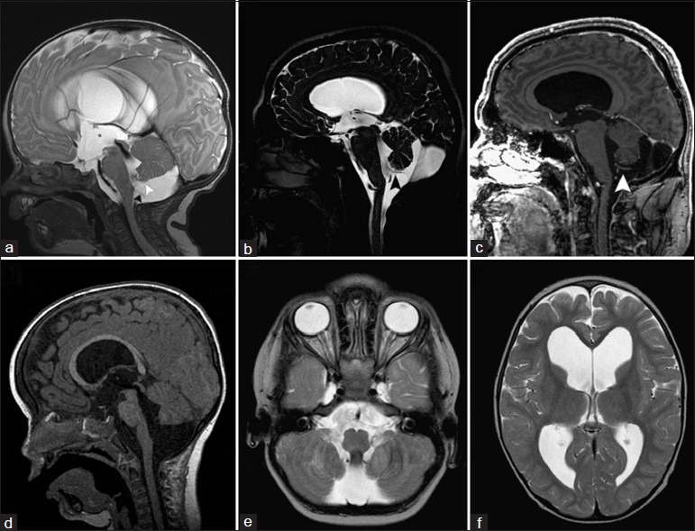
Review
. 2014 Jul 24:5:112.
doi: 10.4103/2152-7806.137533. eCollection 2014.

Blake's pouch cyst

Affiliations
Review

Blake's pouch cyst

Waleed A Azab et al. Surg Neurol Int. .

Abstract

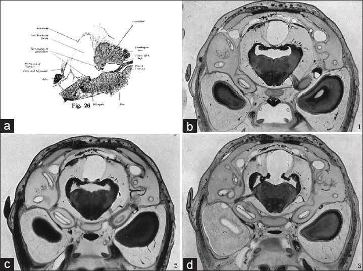
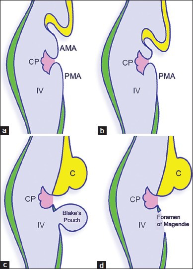
Background: In 1900, Joseph Blake described a transient posterior evagination of the tela choroidea of the fourth ventricle in the normal 130-day old human embryo. He was the first to recognize and fully elucidate on the real nature of the foramen of Magendie as an aperture, which develops within a saccular expansion of the embryonic fourth ventricular cavity. The persistence of this temporary fourth ventricular outpouching into the postnatal period and its significance either as separate entity or as an entity within the Dandy-Walker continuum has over the years been one of the most controversial topics in both neurosurgical and neuroradiological literature.

Methods: A search of the medical literature was conducted for publications addressing the historical, embryological, and neuororadiological features as well as the clinical presentation and management of persistent Blake's pouch.

Results: The literature on the various features of Blake's pouch cyst has limited areas of consensus between various authors.

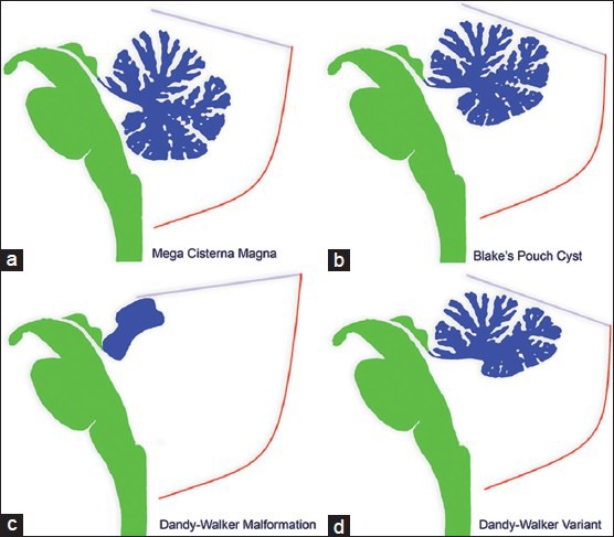
Conclusion: Blake's pouch cyst is a rare entity that is thought to belong to the Dandy-Walker continuum. It has a variable clinical presentation and when symptomatic can be treated with an endoscopic third ventriculostomy or shunting.

Keywords: Blake's pouch cyst; Dandy–Walker continuum; endoscopic; ventriculostomy.


Figures

Figure 1
Figure 1
Joseph A. Blake (1864-1937). Images from the History of Medicine (IHM), National Library of Medicine, History of Medicine Division, Bethesda, Maryland, USA
Figure 2
Figure 2
(a) Image No. 26 from Blake's original work[7] in 1900 demonstrating a sagittal section near the midline in 130 - day human embryo. Note the posterior outpouching of the fourth ventricle. (b-d) Serial sections of the hindbrain of the human embryo at age of 5 months (129 mm crown rump length appearing in Wilson's paper of 1937[35] in support of Blake's observations. The sections are just in front of (b), at the rostral lip (c), and through the anterior part (d) of the foramen of Magendie. Reproduced with permission of John Wiley and Sons, Inc
Figure 3
Figure 3
Embryonic sequence of events in the development of the roof of the fourth ventricle. The plica choroidea (choroid plexus) divides the roof of the fourth ventricle into an anterior membranous area and a posterior membranous area (a). The cerebellar vermis originates from the anterior membranous area (b), which eventually disappears. Blake's pouch appears as a protrusion of the posterior membranous area of the fourth ventricular roof (c), which later communicates with the subarachnoid space forming the foramen of Magendie (d). AMA: Anterior membranous area, C: Cerebellum, CP: Choroid plexus, IV: Fourth ventricle, PMA: Posterior membranous area. (Illustration by Waleed Azab, MD)
Figure 4
Figure 4
Diagrammatic representation of the cerebellar vermis in various entities within the Dandy–Walker complex. (a) Mega cisterna magna. Normal cerebellum and fourth ventricle. (b) Blake's pouch cyst. The vermis is relatively well-developed and nonrotated along with a cystic dilation of the fourth ventricle (c) Dandy-Walker malformation. Rotated small vermis with abnormal foliation and enlarged posterior fossa with elevation of the tentorium and torcula herophili. (d) Dandy-Walker variant. Partial vermian and cerebellar hypoplasia with a prominent retrocerebellar space. (Illustration by Waleed Azab, MD)
Figure 5
Figure 5
(a) Sagittal T2 - weighted MRI image in a case with Blake's pouch cyst. Note the infracerebellar position of the cyst with compressed nonrotated relatively intact cerebellar vermis. The continuation of the fourth ventricular choroid plexus into the Blake's pouch cyst (white arrowhead) and the thin membrane demarcating the cyst from subarachnoid space (black arrowhead) are evident. (b, c) Images from one case reported by Cornips et al.2011[10] demonstrating similar findings. (d-f) Another case from Cornips et al. 2011;[10] (d) Preoperative sagittal T1 - weighted MR image demonstrating marked hydrocephalus with bulging third ventricular floor, downward bending mammillary bodies, and an open aqueduct. The cerebellum is not rotated. (e) Preoperative axial T2 - weighted MR image demonstrating a bilateral indentation on the caudomedial cerebellar surface. (f) Preoperative axial T2 - weighted MR image demonstrating enlarged lateral ventricles without periventricular hyperintensities

Similar articles

Cited by

References

    1. Altman NR, Naidich TP, Braffman BH. Posterior fossa malformations. AJNR Am J Roentgenol. 1992;13:691–724. - PMC - PubMed
    1. Arai H, Sato K. Posterior fossa cysts: Clinical, neuroradiological and surgical features. Childs Nerv Syst. 1991;7:156–64. - PubMed
    1. Barkovich AJ, Kjos BO, Norman D, Edwards MS. Revised classification of posterior fossa cysts and cystlike malformations based on the results of multiplanar MR imaging. AJR Am J Roentgenol. 1989;153:1289–300. - PubMed
    1. Barkovich AJ. Congenital malformations of the brain and skull. In: Barkovich AJ, editor. Pediatric Neuroimaging. 2nd ed. New York: Raven Press; 1994. pp. 177–275.
    1. Barkovich AJ. Developmental disorders of the midbrain and hindbrain. Front Neuroanat. 2012;6:7. - PMC - PubMed