. 2014 Jul 18:2:75.
doi: 10.3389/fped.2014.00075. eCollection 2014.

SMAC Mimetic BV6 Enables Sensitization of Resistant Tumor Cells but also Affects Cytokine-Induced Killer (CIK) Cells: A Potential Challenge for Combination Therapy

Affiliations

SMAC Mimetic BV6 Enables Sensitization of Resistant Tumor Cells but also Affects Cytokine-Induced Killer (CIK) Cells: A Potential Challenge for Combination Therapy

Eva Rettinger et al. Front Pediatr. .

Abstract

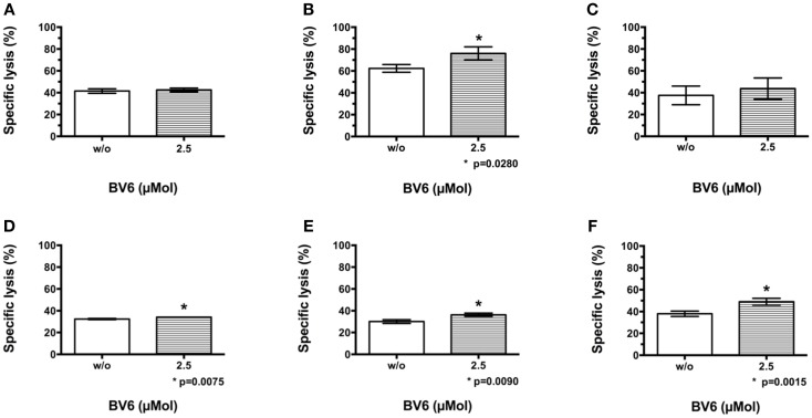
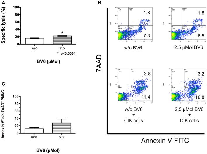
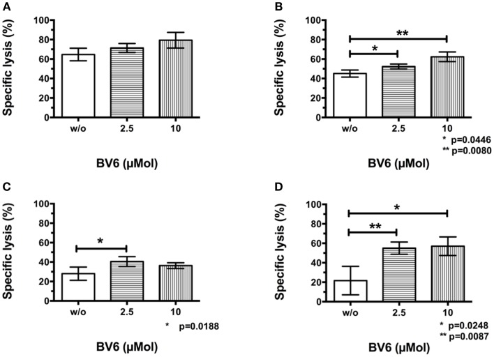
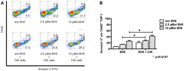
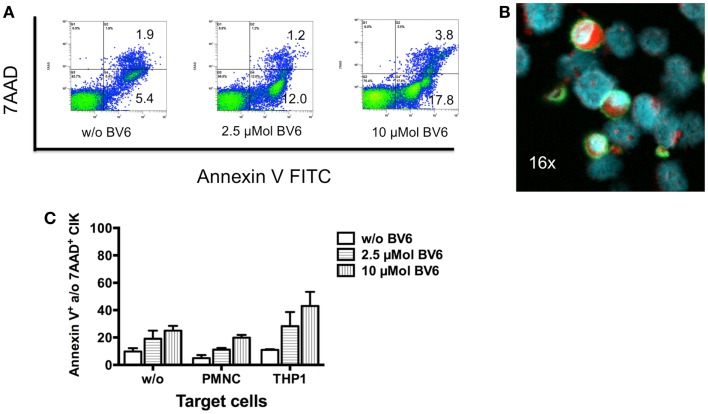
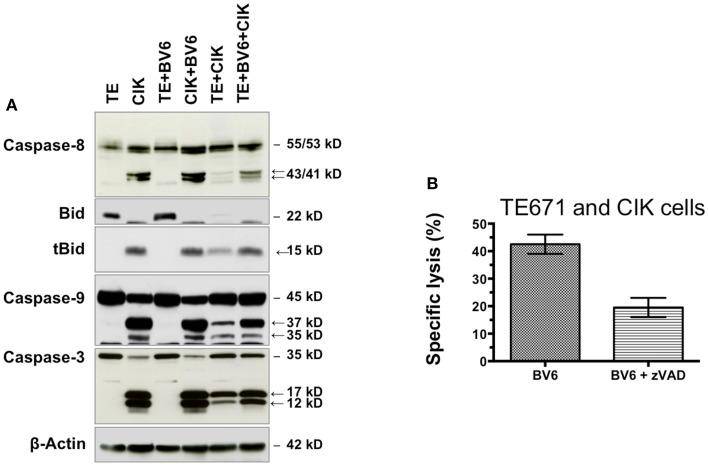
Allogeneic hematopoietic stem cell transplantation (HSCT) is an established treatment option for high-risk hematological malignancies, and may also be offered to patients with solid malignancies refractory to conventional therapies. In case of patients' relapse, refractory tumor cells may then be targeted by cellular therapy-based combination strategies. Here, we investigated the potential of small molecule IAP (SMAC mimetic) BV6 in increasing cytokine-induced killer (CIK) cell-mediated cytotoxicity against different tumor targets. Four-hour pre-incubation with 2.5 μMol BV6 moderately enhanced CIK cell-mediated lysis of hematological (H9, THP-1, and Tanoue) and solid malignancies (RH1, RH30, and TE671). However, BV6 also increased apoptosis of non-malignant cells like peripheral blood mononuclear cells and most notably had an inhibitory effect on immune cells potentially limiting their cytotoxic potential. Hence, cytotoxicity increased in a dose-dependent manner when BV6 was removed before CIK cells were added to tumor targets. However, cytotoxic potential was not further increasable by extending BV6 pre-incubation period of target cells from 4 to 12 h. Molecular studies revealed that BV6 sensitization of target cells involved activation of caspases. Here, we provide evidence that SMAC mimetic may sensitize targets cells for CIK cell-induced cell death. However, BV6 also increased apoptosis of non-malignant cells like CIK cells and peripheral mononuclear cells. These findings may therefore be important for cell- and small molecule IAP-based combination therapies of resistant cancers after allogeneic HSCT.

Keywords: BV6; CIK cells; cellular therapy; leukemia; tumors.


Figures

Figure 1
Figure 1
CIK cell-mediated killing of malignant cells in the presence of BV6. The potential of BV6 to increase CIK cell-mediated killing of hematological (H9, T cell lymphoma (A); THP-1, subtype M4 acute myeloid leukemia (B); Tanoue, precursor-B acute lymphoblastic leukemia (C)) and solid tumor targets (RH1, Ewing sarcoma (D); RH30, alveolar rhabdomyosarcoma (E); TE671, embryonal rhabdomyosarcoma (F)) was analyzed by europium release assay. Therefore, target cells were incubated with 2.5 μMol BV6 for 4 h or remained untreated. Thereafter, CIK cells were added for 3 h at an E:T ratio of 20:1. Results of at least three independent experiments are shown as mean with standard error of mean (mean ± SEM). Treatment with CIK cells alone served as standard control. Combination treatment of CIK cells and BV6 resulted in slightly increased lysis of targets cells in the presence of 2.5 μMol BV6.
Figure 2
Figure 2
CIK cell-mediated killing of non-malignant cells in the presence of BV6. Cytotoxic potential of CIK cells combined with BV6 was analyzed against non-malignant cells like peripheral blood mononuclear cells (PMNCs, E:T ratio 20:1) by europium release assay (A). Analysis of four independent experiments demonstrated as means with standard error of mean (mean ± SEM) revealed that combination treatment significantly increased CIK cell-mediated killing of PMNCs. However, cytotoxicity against non-malignant targets remained relatively low. Accordingly, PMNCs were treated with 2.5 μMol BV6 for 4 h or remained untreated and were incubated with CIK cells at an E:T ratio of 5:1 for 3 h. For analysis of apoptotic and necrotic cells, cell suspensions were stained with Annexin V FITC and 7AAD, respectively. PMNCs were further identified by forward scatter and excluding CD25-expressing larger CIK cells. One representative example showed that compared to untreated controls PMNCs were not affected by BV6, but by CIK cell treatment and furthermore by combination therapy (B). Cytotoxicity data (Annexin V- and/or 7AAD-positive PMNCs) from three independent experiments show as mean ± SEM indicated that BV6 and CIK cell treatment in combination was also toxic for normal cells (C).
Figure 3
Figure 3
CIK cell cytotoxicity against tumor cells after pre-treatment and removal of BV6. Results of at least three independent experiments performed by europium release assay and shown as means with standard error of mean (mean ± SEM) indicated that removal of BV6 after a target cell pre-incubation period of 4 h increased CIK cell-mediated killing of hematological and solid malignancies like THP-1 (A) and RH30 cells (B) in a dose-dependent manner. Furthermore, europium release measurements were performed after extending BV6 sensitization period from 4 to 12 h to increase CIK cell-mediated cytotoxicity towards more resistant target cells like RH30 (C) and TE671 cell lines (D). Cytotoxicity results showed that CIK cell-mediated killing of RH30 and TE671 cell lines was significantly but dose-independently increased, but there was no further enhancement compared to four-hour per-incubation of target cells with BV6.
Figure 4
Figure 4
CIK cell-mediated cytotoxicity after dose escalation of BV6 pre-treatment. Cytotoxic effects of both, increasing doses of BV6 and CIK cells were analyzed by flow cytometry. One representative example is shown (A). Hereby, THP-1 cells were treated with 2.5 or 10 μMol BV6 for 4 h and were incubated with CIK cells at an E:T ratio of 5:1 for another 3 h. THP-1 and CIK cells were differentiated by forward scatter and CD33-expression. Analysis of apoptotic (Annexin V FITC) and necrotic cells (7AAD) showed that combination therapy increased cytotoxicity against CD33-expressing larger THP-1 cells. Cytotoxicity data (Annexin V- and/or 7AAD-positive THP-1 cells) from three independent experiments shown as mean ± SEM demonstrated that BV6 and CIK cell treatment in combination significantly increased cytolysis of THP-1 cells (B).
Figure 5
Figure 5
Induction of apoptosis in CIK cells by SMAC mimetic or killing of target cells. (A) The cytotoxic potential of 2.5 and 10 μMol of the SMAC mimetic BV6 on CIK cells was analyzed by flow cytometry. One representative example showed that CIK cells were affected by increasing doses of BV6 indicated by escalating levels of apoptotic (Annexin V) and necrotic (7AAD) CIK cells (A). After 3 h of co-incubation with alveolar rhabdomyosarcoma cell line RH30 CIK cells adhered to tumor cells, which was shown by one representative example using fluorescence microscope analysis (magnification; 16×). CIK cells were visualized by surface staining with fluorescein isothiocyanate (FITC)-labeled anti-CD45 antibody (green fluorescence). Nuclei of all cells were stained with DAPI (blue fluorescence). As a sign of ongoing apoptosis, CIK cells being in contact with tumor cells and tumor cells both showed intracellular expression of caspase-3 stained with phycoerythrin (PE) (red fluorescence) (B). Cytotoxicity of BV6 on CIK cells alone and in the presence of non-malignant cells (PMNC) and malignant (THP-1) cells (E:T ratio, 5:1) was analyzed by flow cytometry. CIK cells within co-cultures were identified by forward scatter and CD3-, CD25-, and CD33-expression. Cytotoxicity results (mean ± SEM of Annexin V- and/or 7AAD-positive CIK cells) from three independent experiments showed that cytolysis of CIK cells increased with escalating doses of BV6 especially against malignant targets (C).
Figure 6
Figure 6
BV6 and CIK cells reactivate apoptosis in TE671 cells. TE671 cells (TE) were treated for 4 h with 2.5 μM SMAC mimetic BV6 before BV6 was removed and CIK cells were added for 3 h (lane 6). TE and CIK cells alone (lanes 1 and 2), and after 4 h pre-incubation with 2.5 μM BV6 (lines 3 and 4), as well as TE cells after 3 h treatment with CIK cells (lane 5) served as controls. CIK cells within samples of lanes 5 and 6 were removed, before western blot analysis was performed. Caspase activation and Bid cleavage were analyzed. Cleavage fragments are indicated by arrows. β-Actin served as loading control. CIK cell treatment without (lane 5) or particularly with SMAC mimetic BV6 (lane 6) increased TE killing by activation of the caspase cascade. This was evident from the increased cleavage of caspase-8, -3, and -9 into active cleavage fragments in the presence of BV6 and CIK cells, that is, caspase-8 into active p43 and p41 fragments, caspase-3 into active p17 and p12 fragments and caspase-9 into active p37 and p35 fragments. Moreover, Bid was predominately cleaved into tBid, the activated form of Bid, upon BV6 and CIK cell treatment (A). The addition of zVAD a pan caspase inhibitor reduced CIK cell-mediated killing of TE671 cells by 50% demonstrating that BV6 sensitized target cells towards cytotoxicity of CIK cells by caspase activation (B).

Similar articles

Cited by

References

    1. Rettinger E, Willasch AM, Kreyenberg H, Borkhardt A, Holter W, Kremens B, et al. Preemptive immunotherapy in childhood acute myeloid leukemia for patients showing evidence of mixed chimerism after allogeneic stem cell transplantation. Blood (2011) 118:5681–810.1182/blood-2011-04-348805 - DOI - PubMed
    1. Porter DL, Levine BL, Kalos M, Bagg A, June CH. Chimeric antigen receptor-modified T cells in chronic lymphoid leukemia. N Engl J Med (2011) 365:725–3310.1056/NEJMoa1103849 - DOI - PMC - PubMed
    1. Savoldo B, Ramos CA, Liu E, Mims MP, Keating MJ, Carrum G, et al. CD28 costimulation improves expansion and persistence of chimeric antigen receptor-modified T cells in lymphoma patients. J Clin Invest (2011) 121:1822–610.1172/JCI46110 - DOI - PMC - PubMed
    1. Morgan RA, Dudley ME, Wunderlich JR, Hughes MS, Yang JC, Sherry RM, et al. Cancer regression in patients after transfer of genetically engineered lymphocytes. Science (2006) 314:126–910.1126/science.1129003 - DOI - PMC - PubMed
    1. Kronig H, Hofer K, Conrad H, Guilaume P, Muller J, Schiemann M, et al. Allorestricted T lymphocytes with a high avidity T-cell receptor towards NY-ESO-1 have potent anti-tumor activity. Int J Cancer (2009) 125:649–5510.1002/ijc.24414 - DOI - PubMed