. 2014:2014:124902.
doi: 10.1155/2014/124902. Epub 2014 Jul 3.

Effect of angiotensin II and small GTPase Ras signaling pathway inhibition on early renal changes in a murine model of obstructive nephropathy

Affiliations

Effect of angiotensin II and small GTPase Ras signaling pathway inhibition on early renal changes in a murine model of obstructive nephropathy

Ana B Rodríguez-Peña et al. Biomed Res Int. 2014.

Abstract

Tubulointerstitial fibrosis is a major feature of chronic kidney disease. Unilateral ureteral obstruction (UUO) in rodents leads to the development of renal tubulointerstitial fibrosis consistent with histopathological changes observed in advanced chronic kidney disease in humans. The purpose of this study was to assess the effect of inhibiting angiotensin II receptors or Ras activation on early renal fibrotic changes induced by UUO. Animals either received angiotensin II or underwent UUO. UUO animals received either losartan, atorvastatin, and farnesyl transferase inhibitor (FTI) L-744,832, or chaetomellic acid A (ChA). Levels of activated Ras, phospho-ERK1/2, phospho-Akt, fibronectin, and α-smooth muscle actin were subsequently quantified in renal tissue by ELISA, Western blot, and/or immunohistochemistry. Our results demonstrate that administration of angiotensin II induces activation of the small GTPase Ras/Erk/Akt signaling system, suggesting an involvement of angiotensin II in the early obstruction-induced activation of renal Ras. Furthermore, upstream inhibition of Ras signalling by blocking either angiotensin AT1 type receptor or by inhibiting Ras prenylation (atorvastatin, FTI o ChA) reduced the activation of the Ras/Erk/Akt signaling system and decreased the early fibrotic response in the obstructed kidney. This study points out that pharmacological inhibition of Ras activation may hold promise as a future strategy in the prevention of renal fibrosis.


Figures
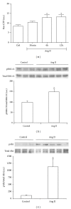
Figure 1
Figure 1
Effect of systemically administrated angiotensin II (Ang II) on Ras signaling pathway. Ras activation was evaluated as Ras GTP by ELISA (a). Phosphorylated (p)-ERK1/2 and p-Akt protein expression were evaluated as the ratio p/total protein by Western blot ((b) and (c), resp.). Bars represent the mean ± SEM of the optical density measured in kidney samples of control saline group (Ctrl; n = 3) and angiotensin II-treated animals (Ang II, 0.8 mg/kg; n = 3–5/per time point). *P < 0.05 versus control group.
Figure 2
Figure 2
Effect of losartan administration on UUO-induced Ras pathway activation and fibrotic changes analyzed by Western blot. Protein expression of Ras (a), ERK1/2 (b), Akt ((c) and (d)), fibronectin (e), and α-SMA (f) detected by immunoblotting. Activation of Ras and ERK1/2 were measured as the ratio phosphorylated (p)/total proteins. Bars represent the mean ± SEM of the optical density measured in nonobstructed (NO) and obstructed (O) kidney samples of saline (n = 3) and losartan- (Los 40 mg/kg; n = 5) treated animals. § P < 0.05 and *Z > 2.6383 versus NO vehicle-treated kidneys of UUO mice.
Figure 3
Figure 3
Effect of losartan, atorvastatin, or farnesyl transferase inhibitor (FTI) administration on renal fibronectin expression detected by immunohistochemistry in UUO mice. Representative interstitial sections from nonobstructed (NO) and obstructed (O) kidneys of UUO untreated control mice ((a) and (b)) and UUO mice treated with losartan ((c) and (d)), atorvastatin ((e) and (f)), or FTI ((g) and (h)). Black bar indicates 100 microns in all panels.
Figure 4
Figure 4
Effect of losartan, atorvastatin, or farnesyl transferase inhibitor (FTI) administration on renal alpha-smooth muscle actin (α-SMA) expression detected by immunohistochemistry in UUO mice. Representative interstitial sections from nonobstructed (NO) and obstructed (O) kidneys of UUO untreated control mice ((a) and (b)) and UUO mice treated with losartan ((c) and (d)), atorvastatin ((e) and (f)) or FTI ((g) and (h)). Black bar indicates 100 microns in all panels.
Figure 5
Figure 5
Effect of atorvastatin administration on UUO-induced Ras pathway activation and fibrotic changes analyzed by Western blot. Protein expression of Ras (a), ERK1/2 (b), Akt ((c) and (d)), fibronectin (e), and alpha-smooth muscle actin (α-SMA) (f) was detected by immunoblotting. Activation of Ras and ERK1/2 was measured as the ratio phosphorylated/total proteins. Bars represent the mean ± SEM of the optical density measured in nonobstructed (NO) and obstructed (O) kidney samples of vehicle (n = 3) and atorvastatin-treated animals (Atorv, 70 mg/kg; n = 4). § P < 0.05 and *Z > 1.9600 versus NO vehicle-treated kidneys of UUO mice. # P < 0.05 versus O vehicle-treated kidneys.
Figure 6
Figure 6
Effect of farnesyl transferase inhibitor (FTI) administration on UUO-induced Ras pathway activation and fibrotic changes analyzed by Western blot. Protein expression of Ras (a), ERK1/2 (b), Akt ((c) and (d)), fibronectin (e), and alpha-smooth muscle actin (α-SMA) (f) was detected by immunoblotting. Activation of Ras and ERK1/2 was measured as the ratio phosphorylated/total proteins. Bars represent the mean ± SEM of the optical density measured in nonobstructed (NO) and obstructed (O) kidney samples of vehicle (n = 3) and FTI-treated animals (40 mg/kg; n = 5). § P < 0.05 and *Z > 2.6383 versus NO vehicle-treated kidneys of UUO mice. # P < 0.05 versus O vehicle-treated kidneys.
Figure 7
Figure 7
Effect of chaetomellic acid A administration on UUO-induced Ras pathway activation and fibrotic changes analyzed by Western blot. Protein expression of pERK1/2 and ERK1/2 (a), pAkt and Akt (b), alpha-smooth muscle actin (α-SMA) (c), and fibronectin (d) was detected by immunoblotting. Bars represent the mean ± SEM of the optical density measured in nonobstructed (NO) and obstructed (O) kidney samples of vehicle (n = 4) and chaetomellic acid A-treated animals (Chaet.; 1.5 mg/kg; n = 4). *P < 0.01 versus NO vehicle-treated kidneys of UUO mice. # P < 0.01 versus O vehicle-treated kidneys. & P < 0.01 versus NO Chaet-treated kidney.
Figure 8
Figure 8
Effect of chaetomellic acid A administration on renal alpha-smooth muscle actin (α-SMA) ((a)–(d)) and fibronectin ((e)–(h)) expression detected by immunohistochemistry in UUO mice. Representative cortical interstitial sections from nonobstructed (NO) and obstructed (O) kidneys of UUO mice treated with vehicle (Control) or chaetomellic acid A (Chaet Acid). Black bar indicates 100 microns in all panels.

Similar articles

Cited by

References

    1. Ucero AC, Benito-Martin A, Izquierdo MC, et al. Unilateral ureteral obstruction: beyond obstruction. International Urology and Nephrology. 2014;46(4):765–776. - PubMed
    1. Bascands J, Schanstra JP. Obstructive nephropathy: insights from genetically engineered animals. Kidney International. 2005;68(3):925–937. - PMC - PubMed
    1. Klahr S, Ishidoya S, Morrissey J. Role of angiotensin II in the tubulointerstitial fibrosis of obstructive nephropathy. American Journal of Kidney Diseases. 1995;26(1):141–146. - PubMed
    1. Pimentel JL, Jr., Montero A, Wang S, Yosipiv I, El-Dahr S, Martinez-Maldonado M. Sequential changes in renal expression of renin-angiotensin system genes in acute unilateral ureteral obstruction. Kidney International. 1995;48(4):1247–1253. - PubMed
    1. Pimentel JL, Jr., Sundell CL, Wang S, Kopp JB, Montero Á, Martínez-Maldonado M. Role of angiotensin II in the expression and regulation of transforming growth factor-β in obstructive nephropathy. Kidney International. 1995;48(4):1233–1246. - PubMed

Publication types

MeSH terms