

Detection of single umbilical artery in the first trimester ultrasound: its value as a marker of fetal malformation
- PMID: 25101287
- PMCID: PMC4101964
- DOI: 10.1155/2014/548729
Detection of single umbilical artery in the first trimester ultrasound: its value as a marker of fetal malformation
Abstract
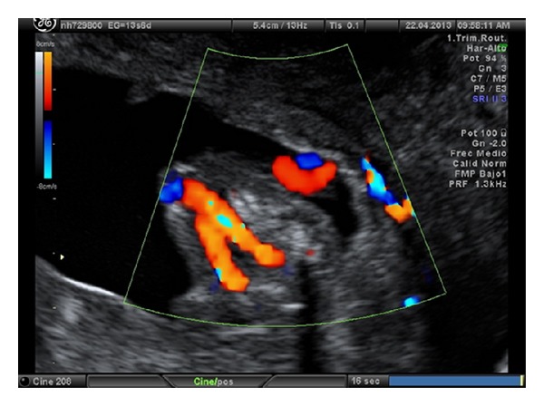
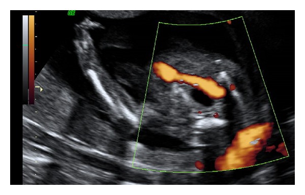
Introduction: The value of a single umbilical artery (SUA) in first trimester ultrasound is not well established. The aim of our study was to determinate the relevance of diagnosis of single umbilical artery in first trimester ultrasound as an early marker suggesting the presence of malformations or associated chromosomopathies.
Material and methods: Retrospective study of clinical cases of SUA diagnosed at the University Hospital Puerta de Hierro in Madrid (Spain) during the first trimester ultrasound between September 2008 and September 2012.
Results: Prevalence of SUA was 1.1% in single pregnancies and 3.3% in twin pregnancies. Sensitivity, specificity, false positive rate, and false negative rate for the finding in the first trimester were 84.2, 99.8, 0.2, and 15.7%, respectively. 17.6% of cases had associated malformations. With an ultrasound in the 16th week most of the cases with significant fetal malformation were diagnosed.
Discussion: SUA is a useful marker in the first trimester for fetal malformation pathology, as it will allow detecting a large number of cases with malformations before 20 weeks of gestation.
Figures
Similar articles
-
Diagnosis of single umbilical artery and risk of foetal congenital malformations by prenatal ultrasound: a retrospective study.BMC Pregnancy Childbirth. 2024 Mar 12;24(1):193. doi: 10.1186/s12884-024-06375-5. BMC Pregnancy Childbirth. 2024. PMID: 38475732 Free PMC article.
-
Agreement between prenatal ultrasonography and fetal autopsy findings: a retrospective study of second trimester terminations of pregnancy.Ultraschall Med. 2012 Dec;33(7):E31-E37. doi: 10.1055/s-0032-1325538. Epub 2012 Nov 29. Ultraschall Med. 2012. PMID: 23196921
-
Twin pregnancies discordant for single umbilical artery.J Reprod Med. 2013 May-Jun;58(5-6):241-5. J Reprod Med. 2013. PMID: 23763010
-
Ultrasound in twin pregnancies.J Obstet Gynaecol Can. 2011 Jun;33(6):643-656. doi: 10.1016/S1701-2163(16)34916-7. J Obstet Gynaecol Can. 2011. PMID: 21846456 Review.
-
Ultrasound diagnosis of structural abnormalities in the first trimester.Prenat Diagn. 2002 Apr;22(4):316-20. doi: 10.1002/pd.309. Prenat Diagn. 2002. PMID: 11981912 Review.
Cited by
-
Analysis of single umbilical artery with concurrent congenital anomaly: Is it a risk factor for poor prognosis? A cross-sectional study.Int J Reprod Biomed. 2024 Mar 25;22(2):139-148. doi: 10.18502/ijrm.v22i2.15710. eCollection 2024 Feb. Int J Reprod Biomed. 2024. PMID: 38628781 Free PMC article.
-
Prenatal Diagnosis of Single Umbilical Artery: Incidence, Counselling and Management in Indian Scenario.J Obstet Gynaecol India. 2018 Dec;68(6):437-439. doi: 10.1007/s13224-017-1089-x. Epub 2018 Jan 25. J Obstet Gynaecol India. 2018. PMID: 30416268 Free PMC article.
-
Diagnosis of single umbilical artery and risk of foetal congenital malformations by prenatal ultrasound: a retrospective study.BMC Pregnancy Childbirth. 2024 Mar 12;24(1):193. doi: 10.1186/s12884-024-06375-5. BMC Pregnancy Childbirth. 2024. PMID: 38475732 Free PMC article.
References
-
- Hua M, Odibo AO, MacOnes GA, Roehl KA, Crane JP, Cahill AG. Single umbilical artery and its associated findings. Obstetrics and Gynecology. 2010;115(5):930–934. - PubMed
-
- Geipel A, Germer U, Welp T, Schwinger E, Gembruch U. Prenatal diagnosis of single umbilical artery: determination of the absent side, associated anomalies. Doppler findings and perinatal outcome. Ultrasound in Obstetrics and Gynecology. 2000;15(2):114–117. - PubMed
-
- Gornall AS, Kurinczuk JJ, Konje JC. Antenatal detection of a single umbilical artery: does it matter? Prenatal Diagnosis. 2003;23(2):117–123. - PubMed
-
- Martínez-Payo C, Gaitero A, Tamarit I, García-Espantaleón M, Goy EI. Perinatal results following the prenatal ultrasound diagnosis of single umbilical artery. Acta Obstetricia et Gynecologica Scandinavica. 2005;84(11):1068–1074. - PubMed
-
- Lubusky M, Dhaifalah I, Prochazka M, et al. Single umbilical artery and its siding in the second trimester of pregnancy: relation to chromosomal defects. Prenatal Diagnosis. 2007;27(4):327–331. - PubMed
MeSH terms
LinkOut - more resources
Full Text Sources
Other Literature Sources
Medical
