

ADAMTS4 and ADAMTS5 knockout mice are protected from versican but not aggrecan or brevican proteolysis during spinal cord injury
- PMID: 25101296
- PMCID: PMC4101972
- DOI: 10.1155/2014/693746
ADAMTS4 and ADAMTS5 knockout mice are protected from versican but not aggrecan or brevican proteolysis during spinal cord injury
Abstract
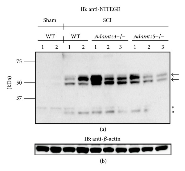
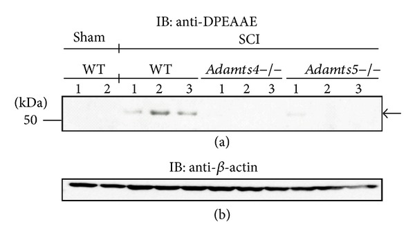
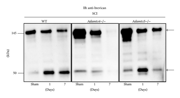
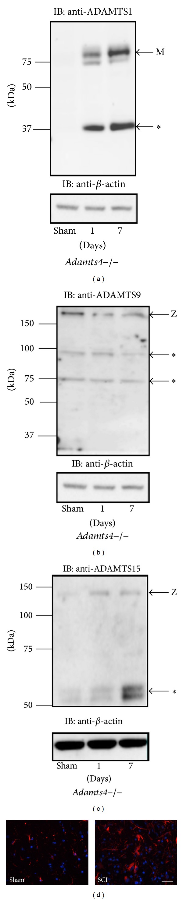
The chondroitin sulfate proteoglycans (CSPGs) aggrecan, versican, and brevican are large aggregating extracellular matrix molecules that inhibit axonal growth of the mature central nervous system (CNS). ADAMTS proteoglycanases, including ADAMTS4 and ADAMTS5, degrade CSPGs, representing potential targets for ameliorating axonal growth-inhibition by CSPG accumulation after CNS injury. We investigated the proteolysis of CSPGs in mice homozygous for Adamts4 or Adamts5 null alleles after spinal cord injury (SCI). ADAMTS-derived 50-60 kDa aggrecan and 50 kDa brevican fragments were observed in Adamts4-/-, Adamts5-/-, and wt mice but not in the sham-operated group. By contrast Adamts4-/- and Adamts5-/- mice were both protected from versican proteolysis with an ADAMTS-generated 70 kDa versican fragment predominately observed in WT mice. ADAMTS1, ADAMTS9, and ADAMTS15 were detected by Western blot in Adamts4-/- mice' spinal cords after SCI. Immunohistochemistry showed astrocyte accumulation at the injury site. These data indicate that aggrecan and brevican proteolysis is compensated in Adamts4-/- or Adamts5-/- mice by ADAMTS proteoglycanase family members but a threshold of versican proteolysis is sensitive to the loss of a single ADAMTS proteoglycanase during SCI. We show robust ADAMTS activity after SCI and exemplify the requirement for collective proteolysis for effective CSPG clearance during SCI.
Figures
References
-
- Fawcett JW, Asher RA. The glial scar and central nervous system repair. Brain Research Bulletin. 1999;49(6):377–391. - PubMed
-
- Iozzo RV, Murdoch AD. Proteoglycans of the extracellular environment: clues from the gene and protein side offer novel perspectives in molecular diversity and function. FASEB Journal. 1996;10(5):598–614. - PubMed
-
- Jäger C, Lendvai D, Seeger G, et al. Perineuronal and perisynaptic extracellular matrix in the human spinal cord. Neuroscience. 2013;238:168–184. - PubMed
-
- Demircan K, Yonezawa T, Takigawa T, et al. ADAMTS1, ADAMTS5, ADAMTS9 and aggrecanase-generated proteoglycan fragments are induced following spinal cord injury in mouse. Neuroscience Letters. 2013;544:25–30. - PubMed
Publication types
MeSH terms
Substances
LinkOut - more resources
Full Text Sources
Other Literature Sources
Medical
Molecular Biology Databases
Miscellaneous
