. 2014 Dec;22(12):2093-2106.
doi: 10.1038/mt.2014.153. Epub 2014 Aug 7.

In vivo evaluation of candidate allele-specific mutant huntingtin gene silencing antisense oligonucleotides

Affiliations

In vivo evaluation of candidate allele-specific mutant huntingtin gene silencing antisense oligonucleotides

Amber L Southwell et al. Mol Ther. 2014 Dec.

Abstract

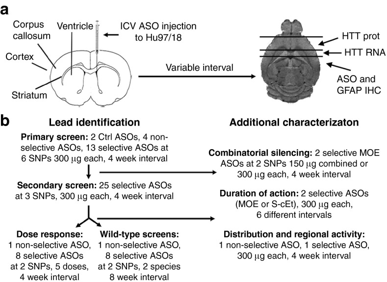
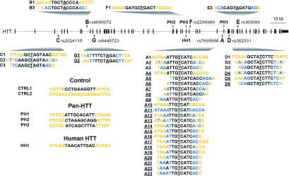
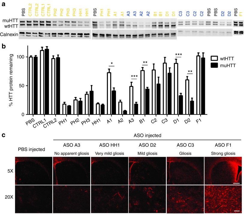
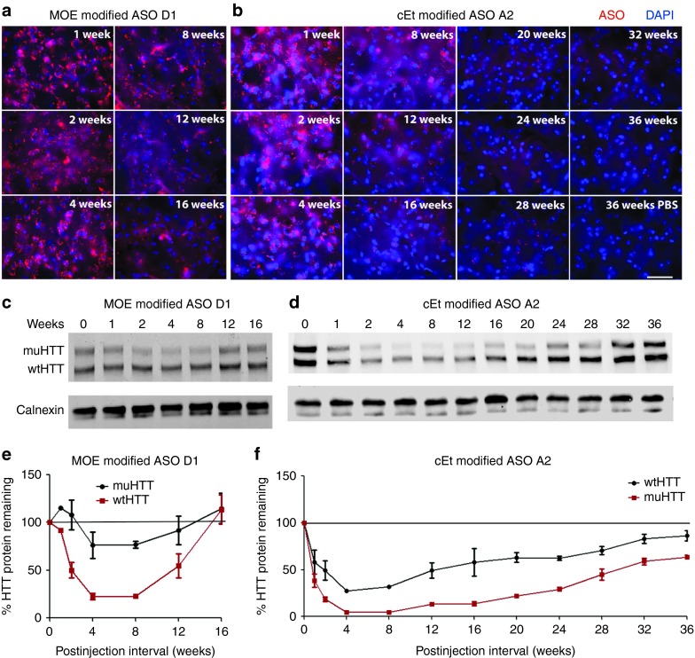
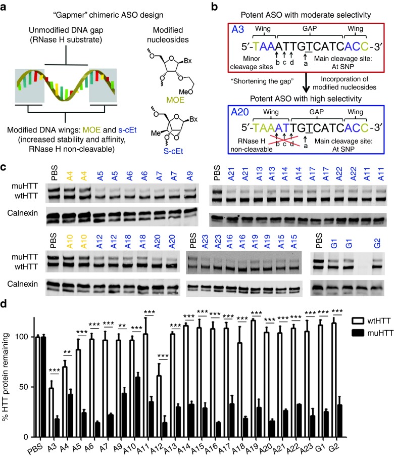
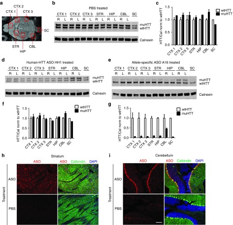
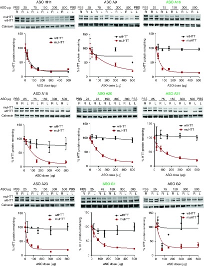
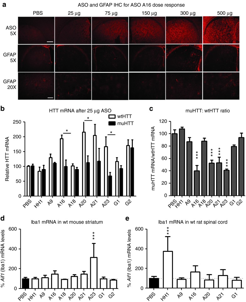
Huntington disease (HD) is a dominant, genetic neurodegenerative disease characterized by progressive loss of voluntary motor control, psychiatric disturbance, and cognitive decline, for which there is currently no disease-modifying therapy. HD is caused by the expansion of a CAG tract in the huntingtin (HTT) gene. The mutant HTT protein (muHTT) acquires toxic functions, and there is significant evidence that muHTT lowering would be therapeutically efficacious. However, the wild-type HTT protein (wtHTT) serves vital functions, making allele-specific muHTT lowering strategies potentially safer than nonselective strategies. CAG tract expansion is associated with single nucleotide polymorphisms (SNPs) that can be targeted by gene silencing reagents such as antisense oligonucleotides (ASOs) to accomplish allele-specific muHTT lowering. Here we evaluate ASOs targeted to HD-associated SNPs in acute in vivo studies including screening, distribution, duration of action and dosing, using a humanized mouse model of HD, Hu97/18, that is heterozygous for the targeted SNPs. We have identified four well-tolerated lead ASOs that potently and selectively silence muHTT at a broad range of doses throughout the central nervous system for 16 weeks or more after a single intracerebroventricular (ICV) injection. With further validation, these ASOs could provide a therapeutic option for individuals afflicted with HD.


Figures

Figure 1
Figure 1
Study overview. (a) Diagram of intracerebroventricular (ICV) antisense oligonucleotides injection and tissue processing method to allow assessment of HTT mRNA and protein and immunohistochemical analyses from the same brains. (b) Study flow chart.
Figure 2
Figure 2
Antisense oligonucleotides (ASOs) in the HTT locus. Diagram showing the location of each targeted single-nucleotide polymorphism (SNP) in the HTT locus and how each evaluated ASO aligns at the SNP. Black-PS, Orange-MOE, Blue-cEt, Green-FHNA, Red-3′ Phosphonate linkage, Target SNP is underlined. Nonunderlined ASOs are from primary screen, underlined ASOs are from secondary screen. Sequence and chemistry of control (CTRL), pan-huntingtin (PH) and human-huntingtin (HH) specific ASOs is also shown.
Figure 3
Figure 3
Single dose screen of 5-9-5 2′-O-methoxyethyl (MOE) and cEt antisense oligonucleotides (ASOs). 300 µg of ASO was delivered by intracerebroventricular (ICV) bolus injection to the right lateral ventricle of 2–3-month-old Hu97/18 mice. Four weeks later, brains were collected and sectioned in a 2 mm coronal rodent brain matrix. The first section containing mostly olfactory bulb was discarded. The second section, containing anterior cortex and striatum, was used for HTT quantitation by allelic separation immunoblotting. The remaining posterior portion of the brain was used for immunohistochemical evaluation of ASO distribution and tolerability. (a) Example western blots showing wt and muHTT protein. ASOs in orange contain only MOE modifications. ASOs in blue contain cEt modifications. (b) Quantitation of HTT protein in both hemispheres of 4 animals. Density of HTT bands was normalized to calnexin loading control and then expressed as a percentage of the same allele (either wtHTT or muHTT) from brain lysates of PBS injected animals on the same membrane. Error bars are SEM. *P < 0.05, **P < 0.01, ***P < 0.001 difference between wt and muHTT by Bonferroni post hoc analysis following two-way analysis of variance. (c) Example immunohistochemistry demonstrating the range of astrogliosis (GFAP reactivity) observed with screened ASOs, as a measure of tolerability. Scale bars are 250 µm for 5× image and 100 µm for 20× image.
Figure 4
Figure 4
Duration of antisense oligonucleotides (ASO) action. Hu97/18 mice received a single 300 µg intracerebroventricular (ICV) injection of ASO. At the indicated intervals brains were harvested and assayed for HTT protein and for ASO by immunohistochemistry (IHC). (a,b) IHC from the central portion of the striatum showing (a) MOE modified ASO D1 or (b) cEt modified ASO A2 in red with DAPI nuclear stain in blue. Scale bar is 25 µm. (c,d) Example western blots showing wt and muHTT protein. (e,f) Quantitation of HTT protein in both hemispheres of two animals. Density of HTT bands was normalized to calnexin loading control and then expressed as a percentage of the same allele (either wtHTT or muHTT) from brain lysates of phosphate-buffered saline–injected animals from the same interval and on the same membrane. Errors bars are SEM.
Figure 5
Figure 5
Secondary screen of enhanced selectivity antisense oligonucleotides (ASO). (a) Typical design of a “gapmer” ASO with high affinity, non-RHaseH cleavable wings surrounding a PS, RNaseH cleavable gap. (b) One strategy for increasing selectivity of a potent ASO by shortening the gap: replacing PS nucleosides at minor cleavage sites with RNAseH noncleavable nucleosides thus restricting cleavage to the main site at the single-nucleotide polymorphism (SNP) of interest. Modified ASOs were then screened in Hu97/18 mice. (c) Example western blots showing wt and muHTT protein. ASOs in orange contain only 5-9-5 2′-O-methoxyethyl modifications. ASOs in blue contain cEt modifications. (d) Quantitation of HTT protein in both hemispheres of four animals. Errors bars are SEM. **P < 0.01, ***P < 0.001 difference between wt and muHTT by Bonferroni post hoc analysis following two-way analysis of variance. Data from the primary screen for the parent molecule, A3, are included on the graph to allow direct comparison.
Figure 6
Figure 6
A single intracerebroventricular (ICV) injection of antisense oligonucleotides (ASO) induces HTT silencing throughout the central nervous system (CNS). Hu97/18 mice received a 300 µg ICV injection of ASO or phosphate-buffered saline (PBS) vehicle. Four weeks later, brains were microdissected into the indicated areas and wt and muHTT were quantified by immunoblot. (a) Diagram of brain areas evaluated. (b) Immunoblot of PBS treated brain demonstrating approximately equivalent wt and muHTT levels in all evaluated regions. (c,f,g) Density of HTT bands is normalized first to calnexin and then to wtHTT density of the same lane. Error bars for bilateral structures are SEM between the two hemispheres. (c) quantitation of HTT in PBS-treated brain. (d) Immunoblot of human-HTT ASO HH1 treated brain showing nonselective reduction of HTT throughout the CNS. (e) Immunoblot of allele-specific ASO A16 treated brain showing selective reduction of muHTT protein throughout the CNS. (f,g) Quantitation of wt and muHTT protein in HH1 and A16 treated brains, respectively. (h,i) immunohistochemistry (IHC) of sagitally sectioned ASO A16 or PBS-injected brain stained for ASO in red, calbindin in green, and 4',6-diamidino-2-phenylindole (DAPI) in blue. (h) In the striatum, ASO distributes evenly, while (i) in the cerebellum it is concentrated in the Purkinje cell layer. Scale bar is 100 µm.
Figure 7
Figure 7
Lead antisense oligonucleotides (ASO) potently and selectively lower muHTT at a broad range of doses. ASO was delivered by intracerebroventricular (ICV) injection at the indicated doses and brains were processed as in screening. Quantitation of HTT protein was performed in both hemispheres of four animals. Density of HTT bands was normalized to calnexin loading control and then expressed as a percentage of the same allele (either wtHTT or muHTT) from brain lysates of phosphate-buffered saline–injected animals on the same membrane. Error bars are SEM. Lead ASOs that passed all screening and dosing criteria are in green.
Figure 8
Figure 8
Lead antisense oligonucleotides (ASO) identified in secondary screen are tolerated at a broad range of doses and efficacious at very low doses. (a–c) ASO was delivered by intracerebroventricular (ICV) injection into Hu97/18 at the indicated doses and brains were processed as in screening. (a) ASO and GFAP immunohistochemistry (IHC) for lead ASO A16 dose response. Induction of gliosis was not observed even at the highest dose. Scale bars are 250 µm for 5× image and 100 µm for 20× image. (b,c) Quantitation of HTT mRNA after ICV delivery of 25 µg of the indicated ASO. (b) relative levels of allelic HTT mRNA in both hemispheres of four animals. *P < 0.05 difference between wt and muHTT by Bonferroni post hoc analysis following two-way analysis of variance (ANOVA). Error bars are SEM. (b) muHTT:wtHTT mRNA ratio. ***P < 0.001 different from phosphate-buffered saline (PBS)–injected animals by Bonferroni post hoc analysis following two-way ANOVA. Error bars are SEM. (d) ASO was delivered to wild-type mice and assessed for tolerability. Aif1 mRNA levels in striatum. PBS compiled from four studies. Error bars are SD (e) ASO was delivered by repeated IT bolus into rats and assessed for tolerability. Aif1 mRNA levels in lumbar spinal cord. PBS compiled from three studies. Error bars are SD. ***P < 0.001 different from PBS-injected animals by Bonferroni post hoc analysis following one-way ANOVA. Data from A20 reported previously.

Similar articles

Cited by

References

    1. Ross CA, Tabrizi SJ. Huntington's disease: from molecular pathogenesis to clinical treatment. Lancet Neurol. 2011;10:83–98. - PubMed
    1. The Huntington's Disease Collaborative Research Group A novel gene containing a trinucleotide repeat that is expanded and unstable on Huntington's disease chromosomes. Cell. 1993;72:971–983. - PubMed
    1. Zuccato C, Valenza M, Cattaneo E. Molecular mechanisms and potential therapeutical targets in Huntington's disease. Physiol Rev. 2010;90:905–981. - PubMed
    1. Videnovic A. Treatment of Huntington disease. Current Treatment Options in Neurology. 2013. pp. 1–15. - PMC - PubMed
    1. Träger U, Andre R, Lahiri N, Magnusson-Lind A, Weiss A, Grueninger S, McKinnon C, et al. HTT-lowering reverses Huntington's disease immune dysfunction caused by NFκB pathway dysregulation. Brain. 2014;137 Pt 3:819–833. - PMC - PubMed

Publication types

MeSH terms