

Effects of Nano-MnO2 on dopaminergic neurons and the spatial learning capability of rats
- PMID: 25101772
- PMCID: PMC4143840
- DOI: 10.3390/ijerph110807918
Effects of Nano-MnO2 on dopaminergic neurons and the spatial learning capability of rats
Abstract
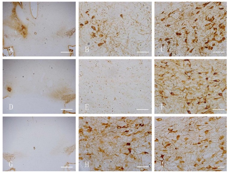
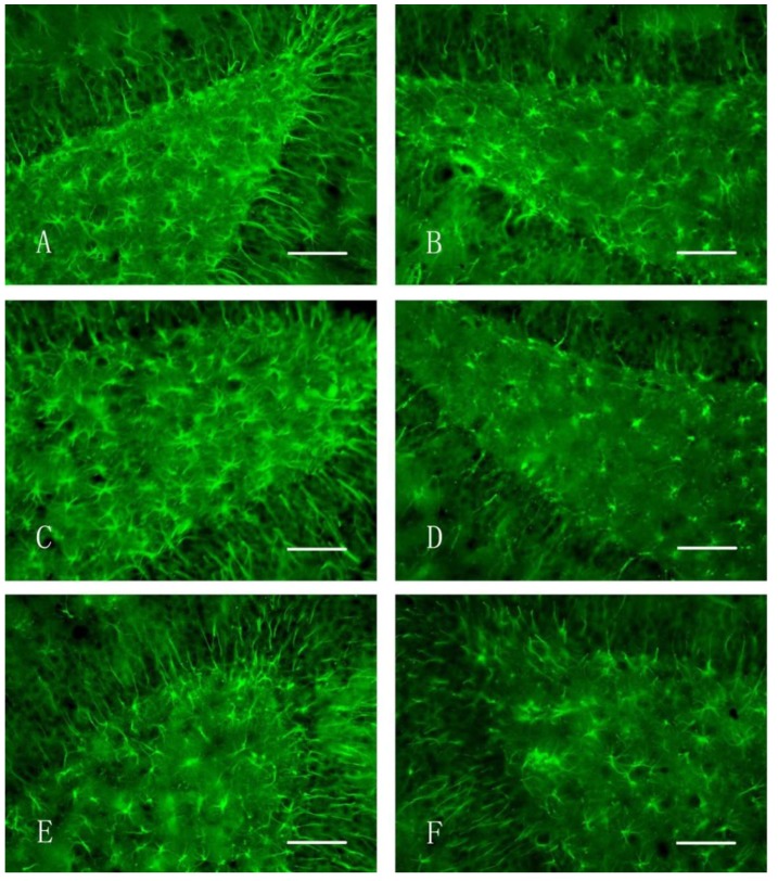
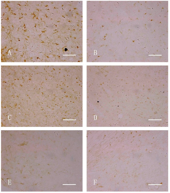
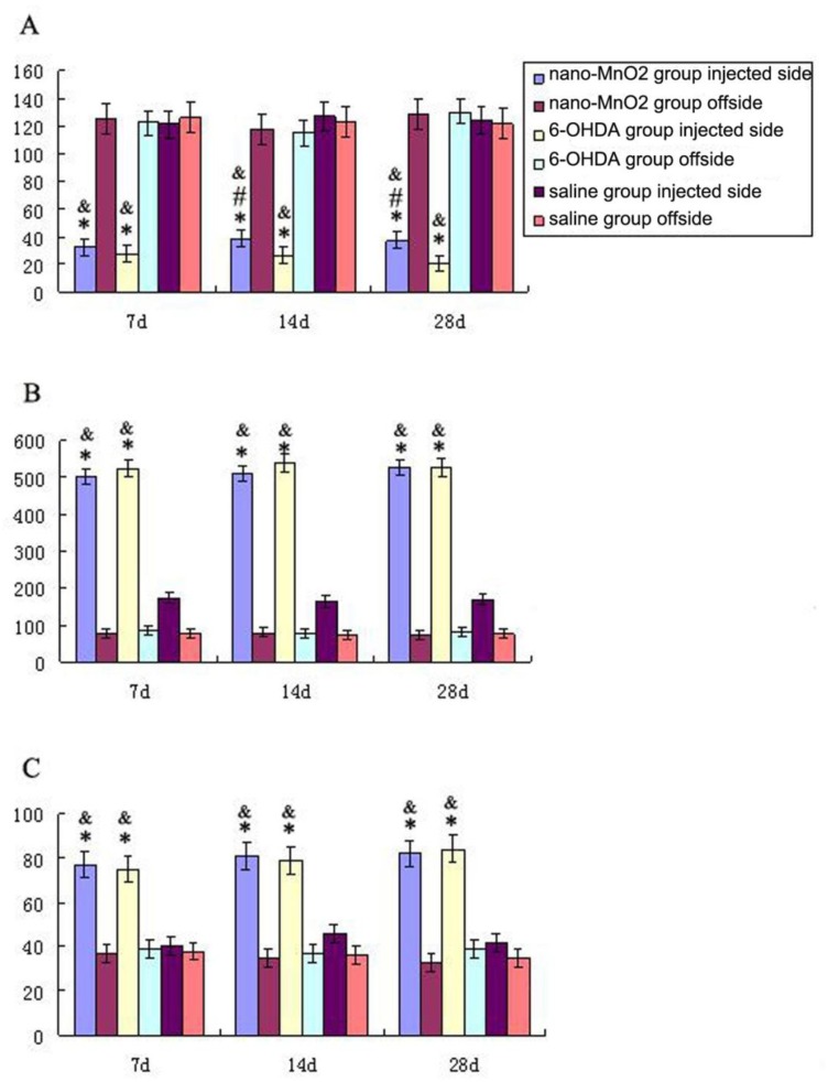
This study aimed to observe the effect of intracerebrally injected nano-MnO2 on neurobehavior and the functions of dopaminergic neurons and astrocytes. Nano-MnO2, 6-OHDA, and saline (control) were injected in the substantia nigra and the ventral tegmental area of Sprague-Dawley rat brains. The neurobehavior of rats was evaluated by Morris water maze test. Tyrosine hydroxylase (TH), inducible nitric oxide synthase (iNOS) and glial fibrillary acidic protein (GFAP) expressions in rat brain were detected by immunohistochemistry. Results showed that the escape latencies of nano-MnO2 treated rat increased significantly compared with control. The number of TH-positive cells decreased, GFAP- and iNOS-positive cells increased significantly in the lesion side of the rat brains compared with the contralateral area in nano-MnO2 group. The same tendencies were observed in nano-MnO2-injected rat brains compared with control. However, in the the positive control, 6-OHDA group, escape latencies increased, TH-positive cell number decreased significantly compared with nano-MnO2 group. The alteration of spatial learning abilities of rats induced by nano-MnO2 may be associated with dopaminergic neuronal dysfunction and astrocyte activation.
Figures
Similar articles
-
Rosiglitazone, a PPAR-γ agonist, protects against striatal dopaminergic neurodegeneration induced by 6-OHDA lesions in the substantia nigra of rats.Toxicol Lett. 2012 Sep 18;213(3):332-44. doi: 10.1016/j.toxlet.2012.07.016. Epub 2012 Jul 27. Toxicol Lett. 2012. PMID: 22842585
-
The acute and the long-term effects of nigral lipopolysaccharide administration on dopaminergic dysfunction and glial cell activation.Eur J Neurosci. 2005 Jul;22(2):317-30. doi: 10.1111/j.1460-9568.2005.04220.x. Eur J Neurosci. 2005. PMID: 16045485
-
[Protective effect of alkaloids from Piper longum in rat dopaminergic neuron injury of 6-OHDA-induced Parkinson's disease].Zhongguo Zhong Yao Za Zhi. 2014 May;39(9):1660-5. Zhongguo Zhong Yao Za Zhi. 2014. PMID: 25095380 Chinese.
-
Dopamine cell morphology and glial cell hypertrophy and process branching in the nigrostriatal system after striatal 6-OHDA analyzed by specific sterological tools.Int J Neurosci. 2005 Apr;115(4):557-82. doi: 10.1080/00207450590521118. Int J Neurosci. 2005. PMID: 15804725
-
Progressive loss of nigrostriatal dopaminergic neurons induced by inflammatory responses to fipronil.Toxicol Lett. 2016 Sep 6;258:36-45. doi: 10.1016/j.toxlet.2016.06.011. Epub 2016 Jun 14. Toxicol Lett. 2016. PMID: 27313094
Cited by
-
Reactive oxygen species-responsive drug delivery systems for the treatment of neurodegenerative diseases.Biomaterials. 2019 Oct;217:119292. doi: 10.1016/j.biomaterials.2019.119292. Epub 2019 Jun 21. Biomaterials. 2019. PMID: 31279098 Free PMC article. Review.
-
Transcriptomics-based investigation of manganese dioxide nanoparticle toxicity in rats' choroid plexus.Sci Rep. 2023 May 25;13(1):8510. doi: 10.1038/s41598-023-35341-y. Sci Rep. 2023. PMID: 37231062 Free PMC article.
-
Application of dental nanomaterials: potential toxicity to the central nervous system.Int J Nanomedicine. 2015 May 14;10:3547-65. doi: 10.2147/IJN.S79892. eCollection 2015. Int J Nanomedicine. 2015. PMID: 25999717 Free PMC article. Review.
-
The effect of iron nanoparticles on performance of cognitive tasks in rats.Environ Sci Pollut Res Int. 2017 Mar;24(9):8700-8710. doi: 10.1007/s11356-017-8531-6. Epub 2017 Feb 16. Environ Sci Pollut Res Int. 2017. PMID: 28210948
-
Silica nanoparticle-exposure during neuronal differentiation modulates dopaminergic and cholinergic phenotypes in SH-SY5Y cells.J Nanobiotechnology. 2019 Apr 1;17(1):46. doi: 10.1186/s12951-019-0482-2. J Nanobiotechnology. 2019. PMID: 30935413 Free PMC article.
References
Publication types
MeSH terms
Substances
LinkOut - more resources
Full Text Sources
Other Literature Sources
Miscellaneous
