. 2014 Nov;26(11):2306-16.
doi: 10.1016/j.cellsig.2014.07.032. Epub 2014 Aug 4.

Hepatocyte growth factor triggers distinct mechanisms of Asef and Tiam1 activation to induce endothelial barrier enhancement

Affiliations

Hepatocyte growth factor triggers distinct mechanisms of Asef and Tiam1 activation to induce endothelial barrier enhancement

Katherine Higginbotham et al. Cell Signal. 2014 Nov.

Abstract

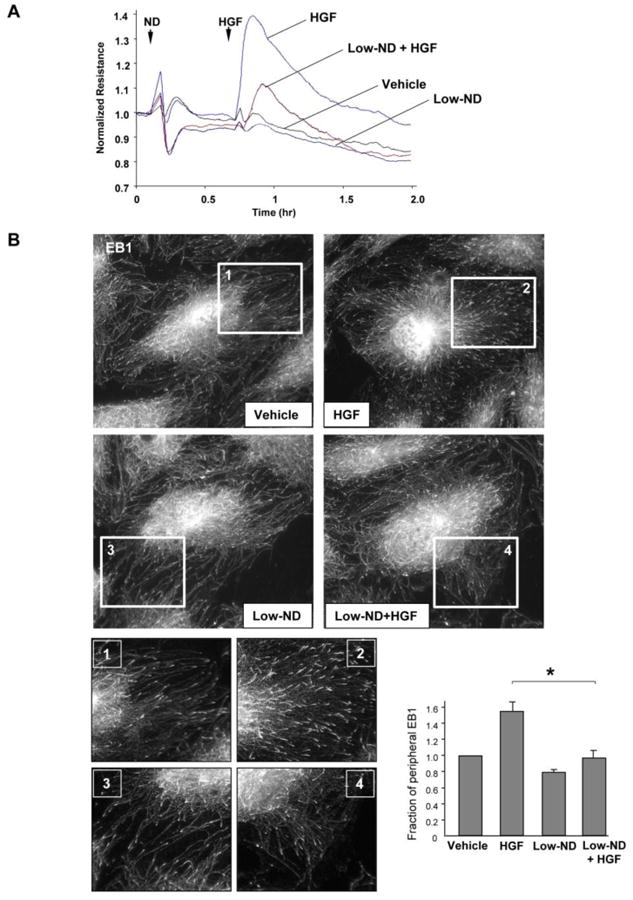
Previous reports described an important role of hepatocyte growth factor (HGF) in mitigation of pulmonary endothelial barrier dysfunction and cell injury induced by pathologic agonists and mechanical forces. HGF protective effects have been associated with Rac-GTPase signaling pathway activated by Rac-specific guanine nucleotide exchange factor Tiam1 and leading to enhancement of intercellular adherens junctions. This study tested involvement of a novel Rac-specific activator, Asef, in endothelial barrier enhancement by HGF and investigated a mechanism of HGF-induced Asef activation. Si-RNA-based knockdown of Tiam1 and Asef had an additive effect on attenuation of HGF-induced Rac activation and endothelial cell (EC) barrier enhancement. Tiam1 and Asef activation was abolished by pharmacologic inhibitors of HGF receptor and PI3-kinase. In contrast to Tiam1, Asef interacted with APC and associated with microtubule fraction upon HGF stimulation. EC treatment by low dose nocodazole to inhibit peripheral microtubule dynamics partially attenuated HGF-induced Asef peripheral translocation, but had negligible effect on Tiam1 translocation. These effects were associated with attenuation of HGF-induced barrier enhancement in EC pretreated with low ND dose and activation of Rac and its cytoskeletal effectors PAK1 and cortactin. These data demonstrate, that in addition to microtubule-independent Tiam1 activation, HGF engages additional microtubule- and APC-dependent pathway of Asef activation. These mechanisms may complement each other to provide the fine tuning of Rac signaling and endothelial barrier enhancement in response to various agonists.

Keywords: Cytoskeleton; Endothelium; Guanine nucleotide exchange factor; HGF; Permeability; Rac GTPase.


Figures

Figure 1
Figure 1. Asef and Tiam1 knockdown attenuates HGF-induced EC barrier enhancement
A - EC grown in 96-well plates were transfected with Asef-specific, Tiam1-specific siRNA or non-specific RNA were stimulated with HGF (50 ng/ml, 10 min). After unbound FITC-avidin was removed, the FITC fluorescence at the bottom of culture dish was measured as described in Methods; *P<0.05 vs. control; **P<0.05 vs. HGF-stimulated EC treated with nonspecific RNA; n=6. B - Pulmonary EC grown on glass coverslips with immobilized biotinylated gelatin (0.25 mg/ml) were transfected with specific siRNA or non-specific RNA. After cell stimulation with vehicle or HGF (50 ng/ml), FITC-avidin (25 μg/ml) was added for 3 min. Unbound FITC-avidin was removed, and FITC fluorescence signal was visualized by fluorescence microscopy.
Figure 2
Figure 2. Role of Asef and Tiam1 in HGF-induced activation of Rac
A – Pulmonary EC were transfected with Asef-specific, Tiam1-specific siRNA, or their combination, or with non-specific RNA and stimulated with HGF (50 ng/ml, 5 min). Rac activation was determined by Rac-GTP pulldown assay. Content of activated Rac was normalized to the total Rac content in EC lysates. siRNA-induced target protein depletion was verified by western blot analysis. Bar graphs represent quantitative densitometry of western blot experiments. *P<0.05 vs. nsRNA; n=3. B – Cells were preincubated with vehicle, c-Met inhibitor (carboxamide 50 nM, 30 min) or PI3-kinase inhibitor (LY294002 20μM, 30 min) followed by stimulation with HGF (5 min). Asef and Tiam1 activation was determined by pulldown assay with immobilized RacG15A and evaluated by increased GEF association with RacG15A. Content of activated Asef or Tiam1 was normalized to the total GEF protein content in EC lysates. Bar graphs represent quantitative densitometry of western blot experiments. *P<0.05 vs. HGF treatment without inhibitors; n=3.
Figure 3
Figure 3. HGF effects on APC, Asef and Tiam1 association with MT fraction and APC-Asef interactions
A – EC were stimulated with HGF (50 ng/ml) followed by isolation of MT-enriched fractionation. APC, Asef and Tiam1 were detected by western blot of MT fractions and normalized to tubulin content. Bar graph represents quantitative densitometry of western blot experiments. *P<0.05; n=3. B – EC were treated with nonspecific and APC-specific siRNA, and HGF-induced accumulation of Asef in MT fraction was assessed. Lower panels show Western blot detection of total Asef and APC protein levels in total cell lysates. Bar graphs represent quantitative densitometry of western blot data. *P<0.05 vs. nsRNA; n=3. C and D - EC pretreated with vehicle, c-Met inhibitor or PI3-kinase inhibitor were stimulated with HGF (50 ng/ml), and APC (C) and Asef (D) proteins were immunoprecipitated under non-denaturing conditions using appropriate antibody. Presence of APC, Asef and Tiam1 in immune complexes was tested by western blot. Results are representative of three to six independent experiments.
Figure 4
Figure 4. Dose-dependent effects of nocodazole on EC permeability and microtubule arrangement
A – EC were treated with the indicated concentrations of nocodazole, and changes in EC permeability were monitored by TER measurements. Shown are representative data from three independent measurements. B – Immunofluorescence staining of microtubule cytoskeleton in EC treated with various nocodazole concentrations was performed using β-tubulin antibody.
Figure 5
Figure 5. Effect of low dose nocodazole on HGF-induced EC barrier enhancement and microtubule peripheral growth
A – Human pulmonary EC were treated with low dose nocodazole (0.05 nM, marked by first arrow). At the time point indicated by second arrow, cells were stimulated with HGF (50 ng/ml), and TER was monitored over 2 hrs. Shown are representative data of four independent experiments. B - EC grown stimulated with HGF with or without pretreatment with low dose nocodazole (0.05 nM) followed by immunostaining with anti-EB1 antibody. Insets show high magnification images of cell peripheral areas with EB1-positive microtubule tips. Bar graph depicts quantitative analysis of peripheral EB1 in methanol-fixed HPAEC monolayers; *P<0.05, n=6.
Figure 6
Figure 6. Effect of low dose nocodazole pretreatment on HGF-induced Asef, Tiam1 and APC peripheral translocation
Human pulmonary EC pretreated with low dose nocodazole (0.05 nM, 15 min) were stimulated with HGF (50 ng/ml). A – Asef, Tiam1 and APC accumulation in membrane/cytoskeletal fraction was monitored by western blot. The content of examined proteins in corresponding total cell lysates was used as a normalization control. Bar graph represents quantitative densitometry of western blot experiments. *P<0.05 vs. HGF without nocodazole treatment; n=4. B - Intracellular redistribution of endogenous Asef an Tiam1 in HGF-stimulated endothelial cells was examined by immunofluorescence staining with appropriate antibody. Shown are representative results of three to five independent experiments.
Figure 7
Figure 7. Pretreatment with low dose nocodazole suppresses HGF-induced activation of Rac pathway and EC barrier enhancement
Human pulmonary EC pretreated with low dose nocodazole (0.05 nM, 15 min) were stimulated with HGF (50 ng/ml). A – Rac activation was assessed by pulldown of Rac-GTP using PAK-PBD beads. B – Rac-dependent PAK and cortactin phosphorylation was assessed by western blot analysis of total cell lysates. C and D – EC permeability was evaluated by XPerT permeability assay described in Methods. After unbound FITC-avidin was removed, the FITC fluorescence signal was visualized by fluorescence microscopy. The bar graph shows quantitative analysis of EC permeability of EC monolayers grown in 96-well plates; *P<0.05 vs. HGF without nocodazole treatment; n=6 (C). Permeability changes were monitored in pulmonary EC grown on glass coverslips with immobilized biotinylated gelatin (0.25 mg/ml) (D).

Similar articles

Cited by

References

    1. Mochizuki N. Circ J. 2009;73(12):2183–2191. - PubMed
    1. Dejana E, Tournier-Lasserve E, Weinstein BM. Dev Cell. 2009;16(2):209–221. - PubMed
    1. Beckers CM, van Hinsbergh VW, van Nieuw Amerongen GP. Thromb Haemost. 2010;103(1):40–55. - PubMed
    1. Tiruppathi C, Minshall RD, Paria BC, Vogel SM, Malik AB. Vascul Pharmacol. 2002;39(4-5):173–185. - PubMed
    1. Hirase T, Node K. Am J Physiol Heart Circ Physiol. 2012;302(3):H499–505. - PubMed

Publication types

MeSH terms