. 2014 Aug 7;9(8):e104045.
doi: 10.1371/journal.pone.0104045. eCollection 2014.

Modulation of PPARγ provides new insights in a stress induced premature senescence model

Affiliations

Modulation of PPARγ provides new insights in a stress induced premature senescence model

Stefania Briganti et al. PLoS One. .

Abstract

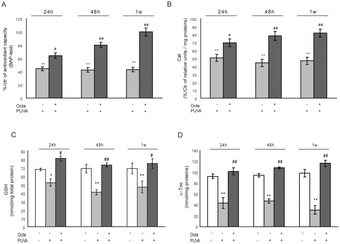
Peroxisome proliferator-activated receptor gamma (PPARγ) may be involved in a key mechanism of the skin aging process, influencing several aspects related to the age-related degeneration of skin cells, including antioxidant unbalance. Therefore, we investigated whether the up-modulation of this nuclear receptor exerts a protective effect in a stress-induced premature senescence (SIPS) model based on a single exposure of human dermal fibroblasts to 8-methoxypsoralen plus + ultraviolet-A-irradiation (PUVA). Among possible PPARγ modulators, we selected 2,4,6-octatrienoic acid (Octa), a member of the parrodiene family, previously reported to promote melanogenesis and antioxidant defense in normal human melanocytes through a mechanism involving PPARγ activation. Exposure to PUVA induced an early and significant decrease in PPARγ expression and activity. PPARγ up-modulation counteracted the antioxidant imbalance induced by PUVA and reduced the expression of stress response genes with a synergistic increase of different components of the cell antioxidant network, such as catalase and reduced glutathione. PUVA-treated fibroblasts grown in the presence of Octa are partially but significantly rescued from the features of the cellular senescence-like phenotype, such as cytoplasmic enlargement, the expression of senescence-associated-β-galactosidase, matrix-metalloproteinase-1, and cell cycle proteins. Moreover, the alterations in the cell membrane lipids, such as the decrease in the polyunsaturated fatty acid content of phospholipids and the increase in cholesterol levels, which are typical features of cell aging, were prevented. Our data suggest that PPARγ is one of the targets of PUVA-SIPS and that its pharmacological up-modulation may represent a novel therapeutic approach for the photooxidative skin damage.


Conflict of interest statement

Competing Interests: The authors received an unrestricted research grant from Giuliani Pharma Milan, thus they declare a financial competing interest. However, the authors confirm that this does not alter their adherence to all PLOS ONE policies on sharing data and materials.

Figures

Figure 1
Figure 1. Evidence for Octa-mediated activation of PPARγ-linked signal transduction.
(A) Activation of RARE. Cells (2×10⁴cells/well) were plated in a 24-well plate and after 24 h they were transfected with RARE. After 24 h, cells were treated with 5µM ReOH for 6 h, 5µM AtRA for 6–48 h, and 2µM Octa for 6–48 h. Measurement of luciferase activity was assessed as reported in Materials and Methods . (B) Quantitative real-time RT-PCR was performed to measure the expression of CRABPII and CYP26A1 mRNA at various time points after treatment with 2µM Octa or 5 µM AtRA. The values were normalized to GAPDH mRNA levels. (C) Quantitative real-time RT-PCR was performed to measure the expression of FABP5 and PPARγ mRNA at various time points after treatment with 2µM Octa or 5µM AtRA. The values were normalized to GAPDH mRNA levels. (D) Luciferase activity analysis of cells transfected with pGL3-(Jwt)3TKLuc reporter construct. After 24 h of transfection, cells were treated with 2µM Octa. The measurement of luciferase activity was carried out 24 h and 48 h after treatment. *p<0.05; **p<0.001 respect to untreated control cells.
Figure 2
Figure 2. Evaluation of PUVA induced effects on PPARγ expression and activity.
(A) Real-time RT-PCR was performed to measure the expression of PPAR-γ mRNA 6 h and 24 h after PUVA exposure. The level of PPAR-γ mRNA was normalized to the expression of GAPDH and is expressed relative to untreated control cells (*p<0.05 respect to Ctr). (B) Luciferase activity analysis of cells transfected with pGL3-(Jwt)3TKLuc reporter construct. After 24 h of transfection, cells were treated with PUVA. The measurement of luciferase activity was carried out 24 h and 48 h after treatment (*p<0.05 respect to Ctr). (C) Real-time RT-PCR was performed to measure the effect of Octa post-treatment on the expression of PPAR-γ mRNA 6 h and 24 h after PUVA exposure. The level of PPAR-γ mRNA was normalized to the expression of GAPDH and is expressed relative to untreated control cells (*p<0.05 respect to Ctr; #p<0.05 respect to PUVA). (D) Luciferase activity analysis of cells transfected with pGL3-(Jwt) 3TKLuc reporter construct. After 24 h of transfection, cells were treated with PUVA and post-incubated with Octa. The measurement of luciferase activity was carried out 24 h and 48 h after treatment (*p<0.05 respect to Ctr; #p<0.05 respect to PUVA).
Figure 3
Figure 3. Effects of PPARγ modulation against PUVA-induced intracellular ROS accumulation and mitochondria damage.
HDFs were treated with PUVA or left untreated (Ctr). Immediately after irradiation PBS was replaced by fresh medium with or without Octa 2µM for 24 h, 48 h or 1 week. (A) Intracellular oxidative stress was assessed by Flow cytometry using the fluorescent probe DCFH2-DA. The median value of fluorescence was used to evaluate the intracellular content of DCF as a measure of the ROS formation. (B) ΔΨm was assessed in live HDFs using the lipophilic cationic probe JC-1. For quantitative fluorescence measurements, cells were rinsed once after JC-1 staining and scanned with a Flow cytometer **p<0.001 statistically different from unirradiated cells; ##p<0.001 compared with PUVA-treated fibroblasts.
Figure 4
Figure 4. Protective action of PPARγ modulation on PUVA-induced imbalance of cell antioxidant system.
HDFs (1×10⁶) were lysed in PBS and protease inhibitor cocktail. Cell lysates were used for analytical determinations. (A) Total antioxidant capacity (TAC) was assessed by BAP-test as described under Materials and Methods section. (B) Cat enzyme activity was determined by spectrophotometry as described under Materials and Methods . (C) GSH concentrations were determined by HPLC-MS as described in Materials and Methods . (D) α-Toc is measured by GC-MS as described in Materials and Methods . *p<0.05; **p<0.001 respect to control fibroblasts; #p<0.05; ##p<0.001 compared with PUVA-treated fibroblasts.
Figure 5
Figure 5. Evidence for PPARγ-induced promotion of cell antioxidant defence.
(A) Octa treatment for 24 h, 48 h and 1 week determined a significant increase of antioxidant cell response. TAC was assessed by BAP-test and Cat enzyme activity was determined by spectrophotometry as described under Materials and Methods section. (B) HDFs were transfected with siRNA specific for PPARγ (siPPARγ) or non-specific siRNA (siCtr). PPARγ level was evaluated by real-time RT-PCR (C) The activity of Cat was assessed in HDFs transfected with siPPARγ or siCtr and exposed to 2µM Octa for 6 h. In parallel Cat activity was measured in HDFs transfected with siPPARγ or siCtr and exposed to PUVA w/o post-incubation with 2µM Octa. *p<0.05; **p<0.001 respect to control fibroblasts; #p<0.05 compared with PUVA-treated fibroblasts.
Figure 6
Figure 6. Possible interference of PPARγ against PUVA induced modulation of the cellular stress response system.
(A) RT-PCR was performed to measure the expression of NRF2 mRNA 6 and 24 h after PUVA exposure, w/o Octa post-incubation. The level of NRF2 mRNA was normalized to the expression of GAPDH and is expressed relative to untreated control cells (**p<0.001 respect to Ctr; ##p<0.001 compared with PUVA-treated fibroblasts). (B) RT-PCR was performed to measure the expression of HO-1 mRNA 6 and 24 h after PUVA exposure, w/o Octa post-incubation. The level of HO-1 mRNA was normalized to the expression of GAPDH and is expressed relative to untreated control cells (**p<0.001 respect to Ctr; ##p<0.001 compared with PUVA-treated fibroblasts). (C) GSH concentrations were determined by HPLC-MS) as described in Materials and Methods (*p<0.05 respect to control fibroblasts). (D) RT-PCR was performed to measure the expression of FoxO1 mRNA 6 and 24 h after PUVA exposure, w/o Octa post-incubation. The level of FoxO1 mRNA was normalized to the expression of GAPDH and is expressed relative to untreated control cells. *p<0.05 respect to control fibroblasts; #p<0.05 compared with PUVA-treated fibroblasts.
Figure 7
Figure 7. Effect of PPARγ modulation on PUVA-induced expression of senescence-like phenotype in HDFs.
After PUVA treatment, HDFs were cultured in the absence or in the presence of 2µM Octa. The medium was changed every 3 days to ensure efficient antioxidant capacity. (A) To evaluate fibroblast morphology, 2 weeks after PUVA in the absence or presence of Octa treatment, cells were fixed and stained with Comassie Brilliant Blue. Scale bar 50 µm. (B) SA-β-gal expression was detected as described in Materials and Methods . The inset represents fibroblasts after PUVA-treatment revealing a senescent phenotype with enlarged cytoplasmic morphology and SA-β-gal expression. The number of SA-β-gal positive fibroblasts is shown as mean ± SD of three independent experiments. **p<0.001 as compared with mock treated controls; ##p<0.001 as compared with PUVA-treated fibroblasts. (C) Supernatants were collected from mock-treated fibroblasts, at 24 h, 48 h and 1 week post PUVA-treatment. MMP-1 release was assessed by ELISA-kit. Three independent experiments in each donor (n = 3) were performed to determine specific MMP-1 protein concentrations in the supernatants. **p<0.001 as compared with mock-treated fibroblasts; #p<0.05; ##p<0.001 as compared with PUVA-treated fibroblasts. (D) Total cellular proteins (30µg/lane) were subject to 10% SDS-PAGE. Variation of protein loading was determined by reblotting membrane with an anti-β-tubulin antibody. Western Blot assays are representative of at least three experiments. Increase of p53 and p21 proteins expression is remarkable 24 h after irradiation as well as until 7 days. Octa treatment decreased PUVA-induced expression of p53 protein (at 24 and 48 h) and of its target gene p21 (at 1 week).
Figure 8
Figure 8. Octa counteracts alteration of lipid cell membrane homeostasis in PUVA treated HDFs.
(A) Polyunsaturated fatty acids of membrane phospholipids (Pl-PUFA) in PUVA-treated HDFs were assessed GC-MS as described in Materials and Methods . (B) Chol content was analyzed by GC-MS as described in Materials and Methods . (C) Early lipid peroxidation products were assessed by the spectrophotometric evaluation of conjugated diene levels as described in Materials and Methods . (D) End products of lipid peroxidation were measured according to TBA assay as described in Materials and Methods . (E) and (F) Chol oxidation was evaluated by assessing 7β-OH-CH and 7-keto-CH as described in Materials and Methods . *p<0.05; **p<0.001 respect to control fibroblasts; #p<0.05; ##p<0.001 compared with PUVA-treated fibroblasts.
Figure 9
Figure 9. PPARγ interference with PUVA-induced phosphorylation pathway and NF-κB activation.
Total cellular proteins (30µg/lane) were subject to 10% SDS-PAGE. Variation of protein loading was determined by reblotting membrane with an anti-GADPH antibody. PUVA-treated HDFs showed an increased phosphorylation of p38 (A) and a decreased expression of IkBα (B). Octa post-treament inhibited p38 phosphorylation (A) as well as decrease of IkBα expression (B) 24 h and 48 h after PUVA treatment, respectively. (C) Densitometric scanning of band intensities obtained from two separate experiments performed in each donor were used to quantify change of protein expression (control value taken as 1-fold in each case). *p<0.05; **p<0.001 respect to control fibroblasts; #p<0.05 compared with PUVA-treated fibroblasts.
Figure 10
Figure 10. Summary scheme of possible role of PPARγ modulation in counteracting PUVA-SIPS of HDFs.
PUVA exposure induced intracellular generation of ROS, alteration of mitochondria function, activation of antioxidant stress response and MAPK phosphorylation pathway, dysregulation of membrane lipid metabolism, DNA-oxidative damage and altered expression of cell cycle regulators. PPARγ modulation by Octa may counteract PUVA-induced senescence-like phenotype. Moreover, Octa ability to reduce phospholipid oxidation and oxysterol generation contributes to the reduction of PUVA-induced inflammatory response and redox imbalance.

Similar articles

Cited by

References

    1. Halliday GM (2005) Inflammation, gene mutation and photoimmunosuppression in response to UVR-induced oxidative damage contributes to photocarcinogenesis. Mutat Res 571: 107–120. - PubMed
    1. Bruls WA, Van Weedlden H, Van der Leun JC (1984) Transmission of UV-irradiation through human epidermal layers as a factor influencing the minimal erythema dose. Photochem Photobiol 39: 63–67. - PubMed
    1. El-Domyati M, Attia S, Saleh F, Brown D, Birk DE, et al. (2002) Intrinsic aging vs. photoaging: a comparative histopathological, immunohistochemical, and ultrastructural study of skin. Exp Dermatol 11: 398–405. - PubMed
    1. Yasui H, Sakurai H (2002) Age-dependent generation of reactive oxygen species in the skin of live hairless rats exposed to UVA light. Exp Dermatol 12: 655–661. - PubMed
    1. Cunningham ML, Krinsky NI, Giovanazzi SM, Peak MJ (1985) Superoxide anion is generated from cellular metabolites by solar radiation and its components. Free Radic Biol Med 1: 381–385. - PubMed

Publication types

MeSH terms

LinkOut - more resources